Spatiotemporal Learning with Context-aware Video Tubelets for Ultrasound Video Analysis

Source: ArXiv

AI Quick Summary

This paper proposes a lightweight framework for ultrasound video analysis that enhances spatiotemporal learning by integrating global context with tubelet-based detection. The method outperforms previous approaches in detecting lung consolidation and pleural effusion, demonstrating efficiency and suitability for real-time clinical workflows.

Share: Share on X LinkedIn

Summary

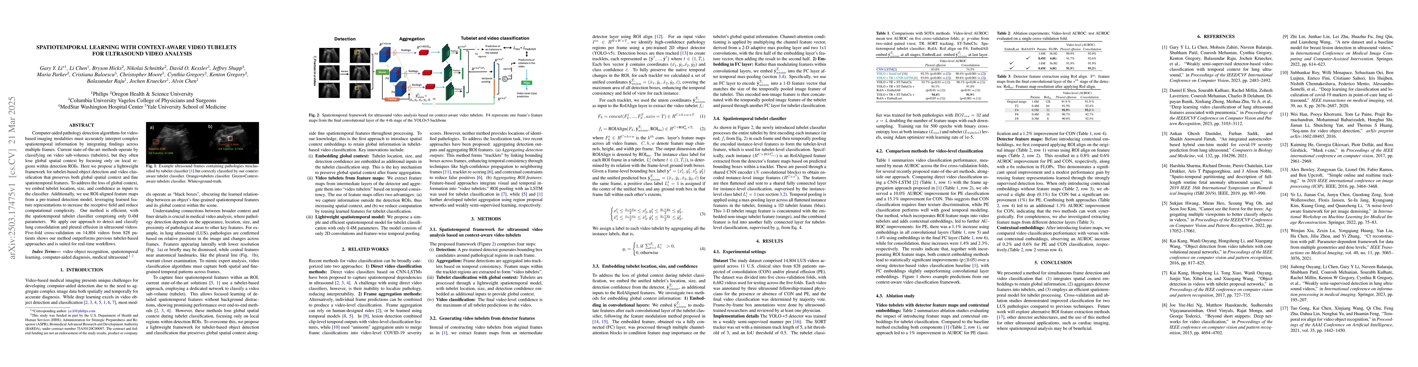
Computer-aided pathology detection algorithms for video-based imaging modalities must accurately interpret complex spatiotemporal information by integrating findings across multiple frames. Current state-of-the-art methods operate by classifying on video sub-volumes (tubelets), but they often lose global spatial context by focusing only on local regions within detection ROIs. Here we propose a lightweight framework for tubelet-based object detection and video classification that preserves both global spatial context and fine spatiotemporal features. To address the loss of global context, we embed tubelet location, size, and confidence as inputs to the classifier. Additionally, we use ROI-aligned feature maps from a pre-trained detection model, leveraging learned feature representations to increase the receptive field and reduce computational complexity. Our method is efficient, with the spatiotemporal tubelet classifier comprising only 0.4M parameters. We apply our approach to detect and classify lung consolidation and pleural effusion in ultrasound videos. Five-fold cross-validation on 14,804 videos from 828 patients shows our method outperforms previous tubelet-based approaches and is suited for real-time workflows.

AI Key Findings

Generated Jun 10, 2025

Methodology

The research proposes a lightweight framework for tubelet-based object detection and video classification in ultrasound video analysis. It preserves global spatial context and fine spatiotemporal features by embedding tubelet location, size, and confidence as inputs to the classifier. ROI-aligned feature maps from a pre-trained detection model are used to increase the receptive field and reduce computational complexity.

Key Results

  • The method outperforms previous tubelet-based approaches in detecting and classifying lung consolidation and pleural effusion in ultrasound videos.
  • Five-fold cross-validation on 14,804 videos from 828 patients demonstrated the superiority of the proposed approach.
  • The spatiotemporal tubelet classifier comprises only 0.4M parameters, making it efficient for real-time workflows.

Significance

This research is significant as it addresses the challenge of accurately interpreting complex spatiotemporal information in video-based imaging modalities, which is crucial for computer-aided pathology detection.

Technical Contribution

The main technical contribution is a novel spatiotemporal framework for ultrasound video analysis that preserves global spatial context and fine spatiotemporal features using context-aware video tubelets.

Novelty

This work introduces the embedding of tubelet location, size, and confidence as additional inputs to the classifier, which is a novel approach to preserving global spatial context in tubelet-based video classification.

Limitations

  • The study is limited to ultrasound video analysis and may not generalize directly to other imaging modalities.
  • Performance evaluation is based on specific pathologies (lung consolidation and pleural effusion) and may not extend to other conditions without further validation.

Future Work

  • Exploration of alternative region of interest (ROI) feature extraction methods.
  • Investigation of the applicability of this method for other ultrasound applications, such as cardiac imaging.

Paper Details

PDF Preview

Download PDF

Citation Network

Current paper (gray), citations (green), references (blue)

Display is limited for performance on very large graphs.

Current Paper
Citations
References
Click to view

No citations found for this paper.

Comments (0)

This website uses cookies to ensure you get the best experience. Learn more