Publication
Metrics
AI Quick Summary
This paper investigates the relationship between the number density of branching vasculature and the scattering properties of soft tissues using speckle. The study finds that the Burr distribution accurately models the histogram of speckle amplitudes, with parameters reflecting the underlying vessel distribution, as demonstrated through both 3D simulations and rat liver scans.
Paper Preview
Abstract
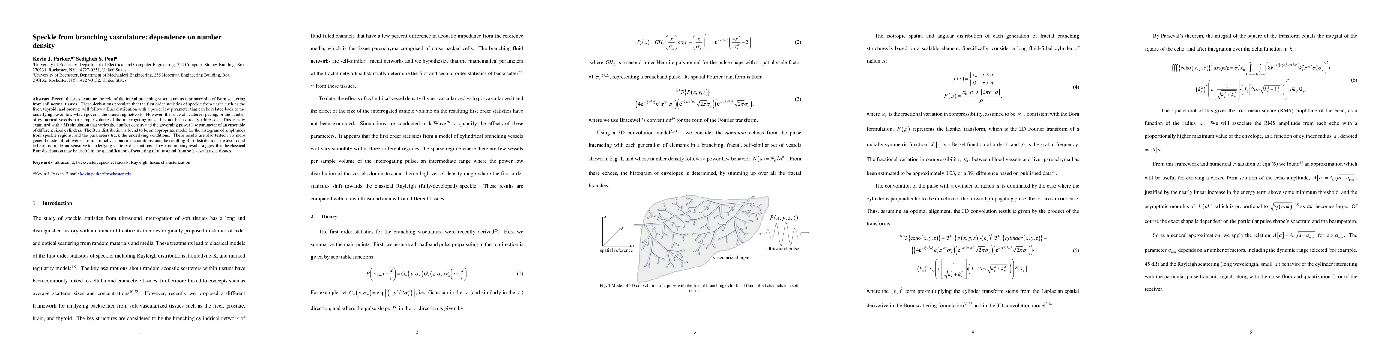
Recent theories examine the role of the fractal branching vasculature as a primary site of Born scattering from soft normal tissues. These derivations postulate that the first order statistics of speckle from tissue such as the liver, thyroid, and prostate will follow a Burr distribution with a power law parameter that can be related back to the underlying power law which governs the branching network. However, the issue of scatterer spacing, or the number of cylindrical vessels per sample volume of the interrogating pulse, has not been directly addressed. This is now examined with a 3D simulation that varies the number density and the governing power law parameter of an ensemble of different sized cylinders. The Burr distribution is found to be an appropriate model for the histogram of amplitudes from speckle regions, and the parameters track the underlying conditions. These results are also tested in a more general model of rat liver scans in normal vs. abnormal conditions, and the resulting Burr distributions are also found to be appropriate and sensitive to underlying scatterer distributions. These preliminary results suggest that the classical Burr distribution may be useful in the quantification of scattering of ultrasound from soft vascularized tissues.
AI Key Findings
Get AI-generated insights about this paper's methodology, results, significance, and more — seven facets brought into focus.
Impact
Paper Details
Authors
PDF Preview
Key Terms
Citation Network
Current paper (gray), citations (green), references (blue)
Display is limited for performance on very large graphs.
Discussion 0