Summary
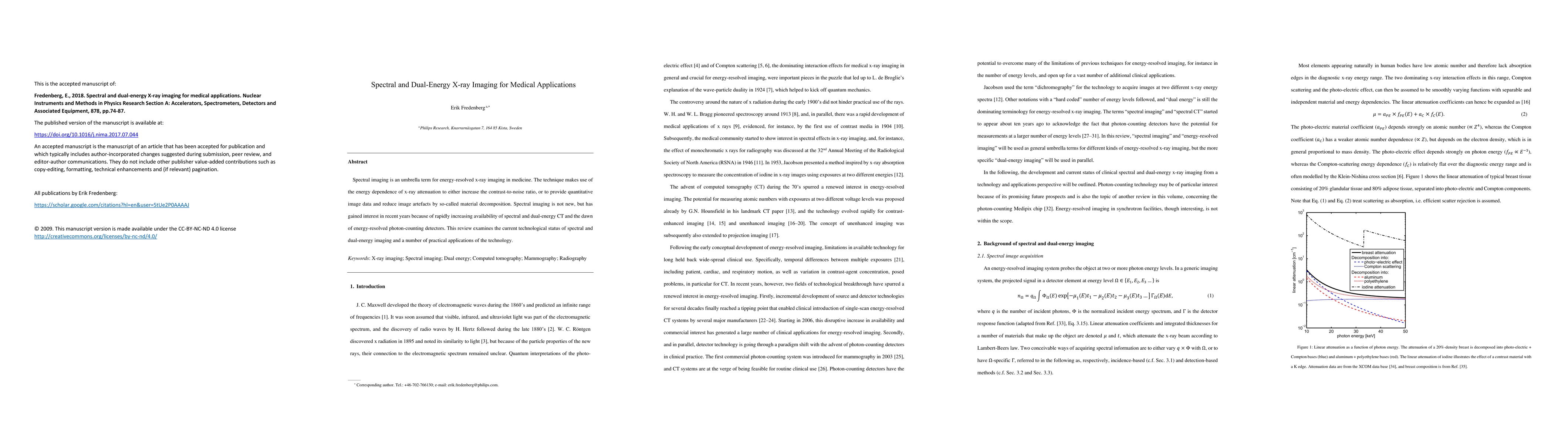
Spectral imaging is an umbrella term for energy-resolved x-ray imaging in medicine. The technique makes use of the energy dependence of x-ray attenuation to either increase the contrast-to-noise ratio, or to provide quantitative image data and reduce image artefacts by so-called material decomposition. Spectral imaging is not new, but has gained interest in recent years because of rapidly increasing availability of spectral and dual-energy CT and the dawn of energy-resolved photon-counting detectors. This review examines the current technological status of spectral and dual-energy imaging and a number of practical applications of the technology.
AI Key Findings
Get AI-generated insights about this paper's methodology, results, and significance.
Paper Details
PDF Preview
Key Terms
Citation Network
Current paper (gray), citations (green), references (blue)
Display is limited for performance on very large graphs.
Similar Papers
Found 4 papers| Title | Authors | Year | Actions |
|---|
Comments (0)