Spectral DiffuserScope: a compact snapshot hyperspectral microscope
Publication
Metrics
Paper Preview
Abstract
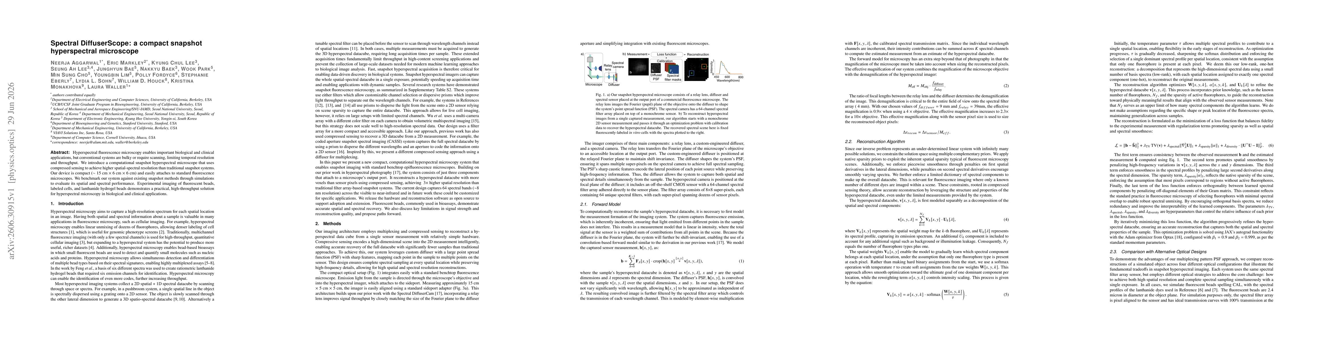
Hyperspectral fluorescence microscopy enables important biological and clinical applications, but conventional systems are bulky or require scanning, limiting temporal resolution and throughput. We introduce a computational snapshot hyperspectral microscope that uses compressed sensing to achieve higher spatial-spectral resolution than traditional snapshot systems. Our device is compact (~15 cm x 6 cm x 6 cm) and easily attaches to standard fluorescence microscopes. We benchmark our system against existing snapshot methods through simulations to evaluate its spatial and spectral performance. Experimental imaging of fluorescent beads, labeled cells, and lanthanide hydrogel beads demonstrates a practical, high-throughput solution for hyperspectral microscopy in biological and clinical applications.
AI Key Findings
Get AI-generated insights about this paper's methodology, results, significance, and more — seven facets brought into focus.
Discussion 0