Speed-of-sound imaging by differential phase contrast with angular compounding
Publication
Metrics
AI Quick Summary
This paper introduces a novel speed-of-sound imaging technique using differential phase contrast with angular compounding, which reveals SoS variations within echogenic samples by monitoring phase shifts from point-like scatterers. The method enhances image sharpness and localization of aberrating inclusions in real time, potentially aiding clinical diagnosis of soft tissue pathologies.
Paper Preview
Abstract
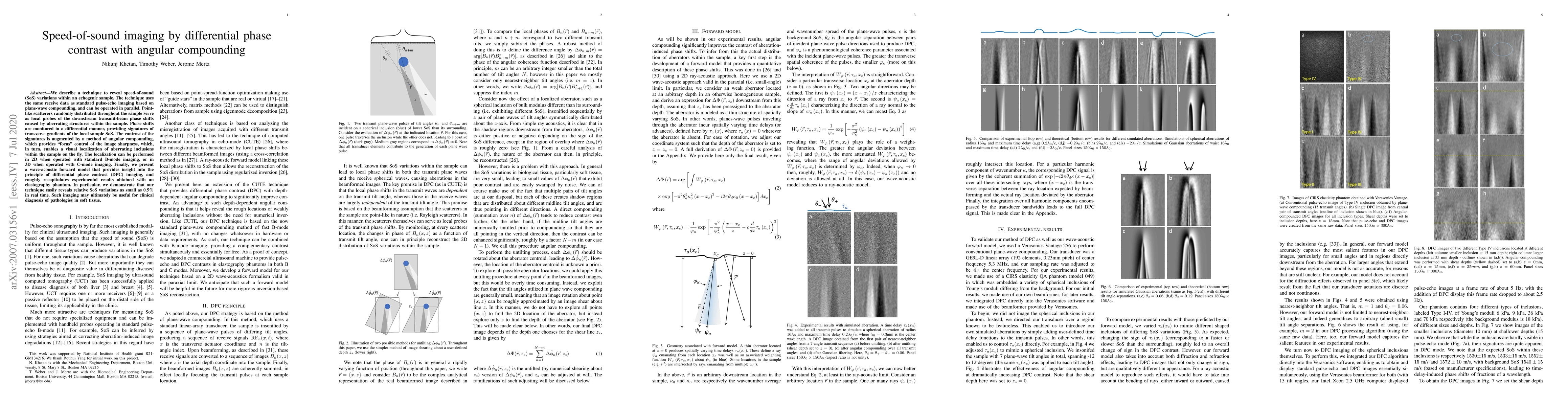
We describe a technique to reveal speed-of-sound (SoS) variations within an echogenic sample. The technique uses the same receive data as standard pulse-echo imaging based on plane-wave compounding, and can be operated in parallel. Point-like scatterers randomly distributed throughout the sample serve as local probes of the downstream transmit-beam phase shifts caused by aberrating structures within the sample. Phase shifts are monitored in a differential manner, providing signatures of transverse gradients of the local sample SoS. The contrast of the signatures is augmented by a method of angular compounding, which provides ``focus" control of the image sharpness, which, in turn, enables a visual localization of aberrating inclusions within the sample on the fly. The localization can be performed in 2D when operated with standard B-mode imaging, or in 3D when operated with C-mode imaging. Finally, we present a wave-acoustic forward model that provides insight into the principle of differential phase contrast (DPC) imaging, and roughly recapitulates experimental results obtained with an elastography phantom. In particular, we demonstrate that our technique easily reveals relative SoS variations as small as 0.5\% in real time. Such imaging may ultimately be useful for clinical diagnosis of pathologies in soft tissue.
AI Key Findings
Get AI-generated insights about this paper's methodology, results, significance, and more — seven facets brought into focus.
Impact
Paper Details
Authors
PDF Preview
Key Terms
Citation Network
Current paper (gray), citations (green), references (blue)
Display is limited for performance on very large graphs.
Discussion 0