Speed-of-Sound Imaging using Diverging Waves
Publication
Metrics
AI Quick Summary
This paper proposes using diverging waves (DW) for speed-of-sound (SoS) imaging to improve reconstruction accuracy over plane wave (PW) insonifications. Results show a 22% reduction in reconstruction error and a 55% improvement in contrast, demonstrating better performance in both simulated and real tissue phantoms.
Paper Preview
Abstract
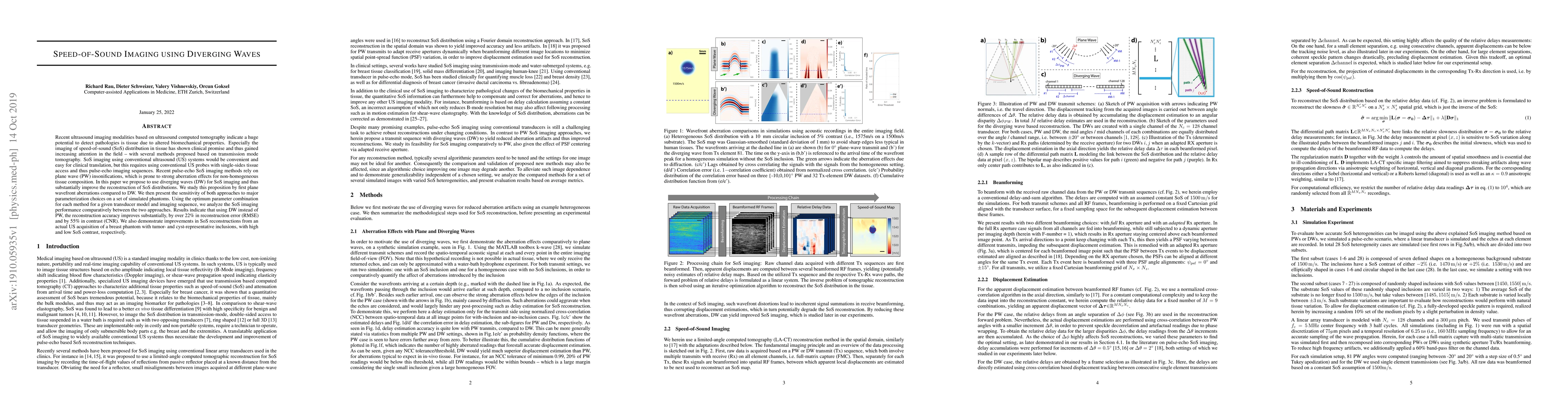
Recent ultrasound imaging modalities based on ultrasound computed tomography indicate a huge potential to detect pathologies is tissue due to altered biomechanical properties. Especially the imaging of speed-of-sound (SoS) distribution in tissue has shown clinical promise and thus gained increasing attention in the field -- with several methods proposed based on transmission mode tomography. SoS imaging using conventional ultrasound (US) systems would be convenient and easy for clinical translation, but this requires using conventional US probes with single-sides tissue access and thus pulse-echo imaging sequences. Recent pulse-echo SoS imaging methods rely on plane wave (PW) insonifications, which is prone to strong aberration effects for non-homogeneous tissue composition. In this paper we propose to use diverging waves (DW) for SoS imaging and thus substantially improve the reconstruction of SoS distributions. We study this proposition by first plane wavefront aberrations compared to DW. We then present the sensitivity of both approaches to major parameterization choices on a set of simulated phantoms. Using the optimum parameter combination for each method for a given transducer model and imaging sequence, we analyze the SoS imaging performance comparatively between the two approaches. Results indicate that using DW instead of PW, the reconstruction accuracy improves substantially, by over 22% in reconstruction error (RMSE) and by 55% in contrast (CNR). We also demonstrate improvements in SoS reconstructions from an actual US acquisition of a breast phantom with tumor- and cyst-representative inclusions, with high and low SoS contrast, respectively.
AI Key Findings
Get AI-generated insights about this paper's methodology, results, significance, and more — seven facets brought into focus.
Impact
Paper Details
Authors
PDF Preview
Key Terms
Citation Network
Current paper (gray), citations (green), references (blue)
Display is limited for performance on very large graphs.
Discussion 0