Summary

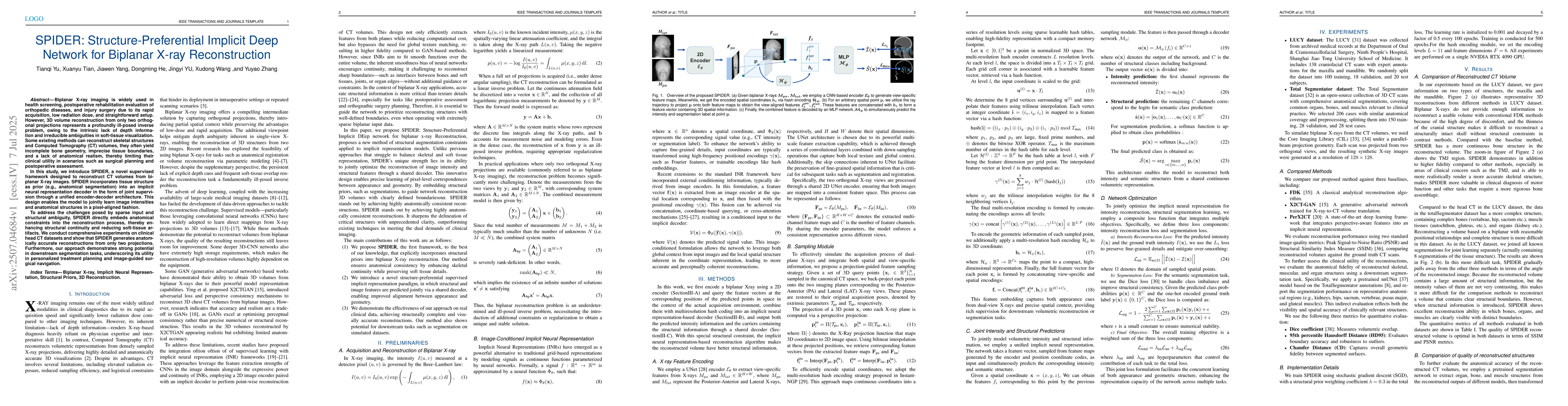
Biplanar X-ray imaging is widely used in health screening, postoperative rehabilitation evaluation of orthopedic diseases, and injury surgery due to its rapid acquisition, low radiation dose, and straightforward setup. However, 3D volume reconstruction from only two orthogonal projections represents a profoundly ill-posed inverse problem, owing to the intrinsic lack of depth information and irreducible ambiguities in soft-tissue visualization. Some existing methods can reconstruct skeletal structures and Computed Tomography (CT) volumes, they often yield incomplete bone geometry, imprecise tissue boundaries, and a lack of anatomical realism, thereby limiting their clinical utility in scenarios such as surgical planning and postoperative assessment. In this study, we introduce SPIDER, a novel supervised framework designed to reconstruct CT volumes from biplanar X-ray images. SPIDER incorporates tissue structure as prior (e.g., anatomical segmentation) into an implicit neural representation decoder in the form of joint supervision through a unified encoder-decoder architecture. This design enables the model to jointly learn image intensities and anatomical structures in a pixel-aligned fashion. To address the challenges posed by sparse input and structural ambiguity, SPIDER directly embeds anatomical constraints into the reconstruction process, thereby enhancing structural continuity and reducing soft-tissue artifacts. We conduct comprehensive experiments on clinical head CT datasets and show that SPIDER generates anatomically accurate reconstructions from only two projections. Furthermore, our approach demonstrates strong potential in downstream segmentation tasks, underscoring its utility in personalized treatment planning and image-guided surgical navigation.

AI Key Findings

Generated Sep 03, 2025

Methodology

The research introduces SPIDER, a structure-preferential implicit deep network for biplanar X-ray reconstruction. It incorporates tissue structure as prior into an implicit neural representation decoder via joint supervision in a unified encoder-decoder architecture, enabling the model to learn image intensities and anatomical structures in a pixel-aligned fashion.

Key Results

  • SPIDER generates anatomically accurate 3D volume reconstructions from only two biplanar X-ray projections.
  • Compared to existing methods, SPIDER demonstrates improved structural continuity and reduced soft-tissue artifacts.
  • The approach shows strong potential in downstream segmentation tasks, enhancing its utility in personalized treatment planning and image-guided surgical navigation.
  • SPIDER outperforms state-of-the-art methods like X2CT-GAN and PerX2CT in both image quality and structural fidelity.
  • The reconstructed volumes from SPIDER exhibit enhanced anatomical continuity and clearer tissue boundaries, especially in clinically important regions such as the temporomandibular joint.

Significance

This research is significant as it addresses the ill-posed inverse problem in biplanar X-ray reconstruction, providing a practical and effective solution for sparse-view medical imaging. The proposed SPIDER framework bridges the gap between data efficiency and clinical fidelity, making it a promising tool for real-world applications like surgical planning and diagnostic support.

Technical Contribution

The paper presents SPIDER, a structure-prior implicit deep network that embeds anatomical structural priors directly into an implicit neural representation decoder through joint training, enabling the decoder to learn the pixel-to-pixel mapping of intensity and structure information.

Novelty

SPIDER differs from existing methods by directly incorporating anatomical structural priors into the implicit neural representation decoder, allowing for joint learning of image intensities and anatomical structures in a pixel-aligned manner. This novel approach results in improved structural fidelity and clinical utility compared to previous methods.

Limitations

  • SPIDER may cause some parts of the reconstruction outside the region of joining structural information to become worse and lack some high-frequency information.
  • The method is sensitive to the weights of the joint training loss function, requiring careful tuning for optimal performance.

Future Work

  • Explore methods to mitigate the degradation of image quality in regions without structural information.
  • Investigate adaptive weighting strategies for the joint training loss function to improve training stability and reconstruction quality.

Paper Details

PDF Preview

Download PDF

Citation Network

Current paper (gray), citations (green), references (blue)

Display is limited for performance on very large graphs.

Current Paper
Citations
References
Click to view

No citations found for this paper.

Comments (0)

This website uses cookies to ensure you get the best experience. Learn more