The study extends vertex models to curved manifolds by placing cells on ellipsoidal surfaces with geodesic cell edges, using a mean-field energy e = sum_i [kA(ai−1)^2 + (pi−p0)^2]/2 and deriving area/perimeter on curved surfaces as functions of local Gaussian curvature κ; cell dynamics follow overdamped motion η drα/dt = Fα with forces from area and perimeter terms, while enforcing uniform tissue stresses across the curved surface to achieve force balance. Analytical approximations for area a and perimeter p on small cells in a κ patch are derived via Riemann normal coordinates, and derivatives ∂a/∂rα and ∂p/∂rα are computed to obtain vertex forces, enabling simulation of confluent tissues on ellipsoids and examination of area and perimeter patterning induced by curvature.
Publication
Metrics
Quick Answers
What methodology did the authors use?
The study extends vertex models to curved manifolds by placing cells on ellipsoidal surfaces with geodesic cell edges, using a mean-field energy e = sum_i [kA(ai−1)^2 + (pi−p0)^2]/2 and deriving area/perimeter on curved surfaces as functions of local Gaussian curvature κ; cell dynamics follow overdamped motion η drα/dt = Fα with forces from area and perimeter terms, while enforcing uniform... More in Methodology →
What are the key results?
On ellipsoidal surfaces, cells near the poles become larger than those near the equator, revealing spontaneous cell-area patterning that strengthens with increasing aspect ratio. — Perimeter patterns arise from the competition between intrinsic geometric tendencies and force-balance deformations, and can invert beyond a critical shape index. More in Key Results →
Why is this work significant?
Demonstrates a physical, curvature-enabled mechanism for spontaneous spatial variation in cell size within confluent tissues, suggesting curvature sensing can emerge from geometry–mechanics interplay without biochemical gradients, with potential relevance to embryogenesis and organogenesis. More in Significance →
What are the main limitations?
Analyses rely on small-curvature expansions and mean-field approximations, potentially limiting accuracy for large cells or high curvature regions. — Model focuses on elastic and tension-driven contributions; active cellular processes or heterogeneity in cell properties are not exhaustively explored. More in Limitations →
Paper Preview
Abstract
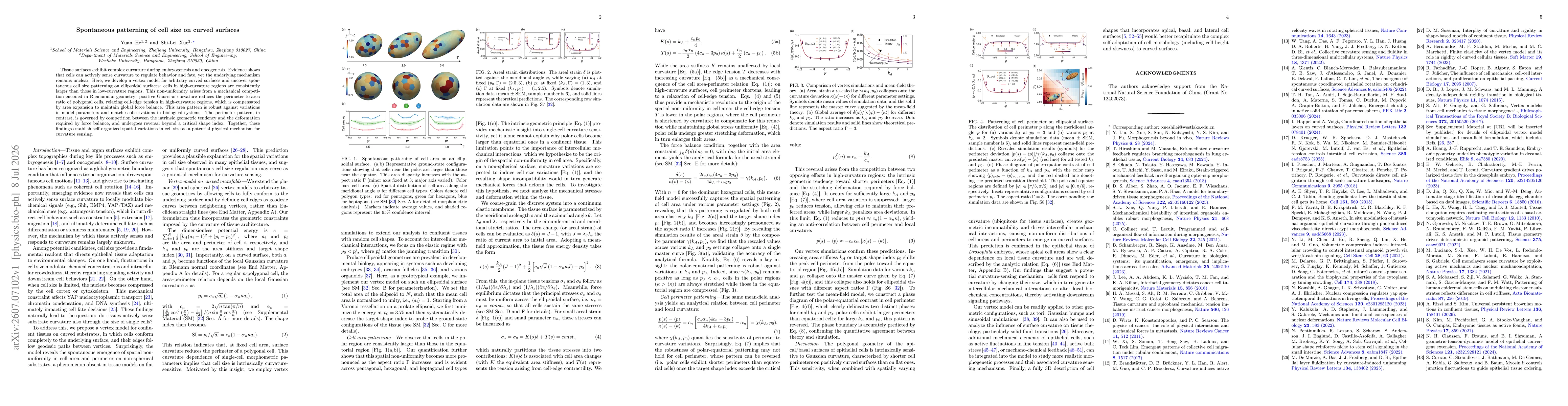
Tissue surfaces exhibit complex curvature during embryogenesis and oncogenesis. Evidence shows that cells can actively sense curvature to regulate behavior and fate, yet the underlying mechanism remains unclear. Here, we develop a vertex model for arbitrary curved surfaces and uncover spontaneous cell size patterning on ellipsoidal surfaces: cells in high-curvature regions are consistently larger than those in low-curvature regions. This non-uniformity arises from a mechanical competition encoded in Riemannian geometry: positive Gaussian curvature reduces the perimeter-to-area ratio of polygonal cells, relaxing cell-edge tension in high-curvature regions, which is compensated by area expansion to maintain global force balance. This area pattern is robust against variations in model parameters and matches observations in biological systems. The perimeter pattern, in contrast, is governed by competition between the intrinsic geometric tendency and the deformation required by force balance, and undergoes reversal beyond a critical shape index. Together, these findings establish self-organized spatial variations in cell size as a potential physical mechanism for curvature sensing.
Key Findings, in focus
Seven facets of this paper, analysed and brought into focus by AI.
Demonstrates a physical, curvature-enabled mechanism for spontaneous spatial variation in cell size within confluent tissues, suggesting curvature sensing can emerge from geometry–mechanics interplay without biochemical gradients, with potential relevance to embryogenesis and organogenesis.
- On ellipsoidal surfaces, cells near the poles become larger than those near the equator, revealing spontaneous cell-area patterning that strengthens with increasing aspect ratio.
- Perimeter patterns arise from the competition between intrinsic geometric tendencies and force-balance deformations, and can invert beyond a critical shape index.
- A continuum elastic description decouples area changes (K(s)δ) from edge-tension contributions (T(s)), explaining how curvature drives spatially varying cell areas while preserving overall force balance.
Demonstrates a physical, curvature-enabled mechanism for spontaneous spatial variation in cell size within confluent tissues, suggesting curvature sensing can emerge from geometry–mechanics interplay without biochemical gradients, with potential relevance to embryogenesis and organogenesis.
Derivation of area and perimeter expressions for polygonal cells on curved surfaces using local Gaussian curvature in Riemann normal coordinates, including explicit derivatives of area and geodesic-edge lengths with respect to vertex positions; integration of these derivatives into an overdamped vertex dynamics framework on curved manifolds.
First demonstration of spontaneous curvature-induced cell-size patterns in a confluent tissue using a curved-geometry vertex model; identification of a curvature-dependent relaxation of edge tension and compensatory area expansion as the mechanism behind pattern formation on non-spherical surfaces.
- Analyses rely on small-curvature expansions and mean-field approximations, potentially limiting accuracy for large cells or high curvature regions.
- Model focuses on elastic and tension-driven contributions; active cellular processes or heterogeneity in cell properties are not exhaustively explored.
- Simulations are performed on idealized ellipsoidal geometries; applicability to irregular in vivo geometries remains to be tested.
- Extend to more complex curved geometries and non-uniform tissue properties to test robustness of area/perimeter patterning.
- Incorporate active remodeling, cell division, and heterogeneous mechanical parameters to assess impact on curvature sensing.
- Direct experimental validation in epithelial tissues with controllable curvature to connect predictions with biological observables.
Discussion 0