SQUID-based instrumentation for ultra-low-field MRI
Publication
Metrics
AI Quick Summary
This paper describes a seven-channel SQUID-based system for ultra-low-field MRI (ULF MRI) and magnetoencephalography (MEG), achieving microtesla-range field resolutions. The system acquired 3D images of a preserved sheep brain at 46 microtesla, demonstrating a resolution of 2.5 mm x 2.5 mm x 5 mm, and compares these results to conventional high-field MRI images.
Paper Preview
Abstract
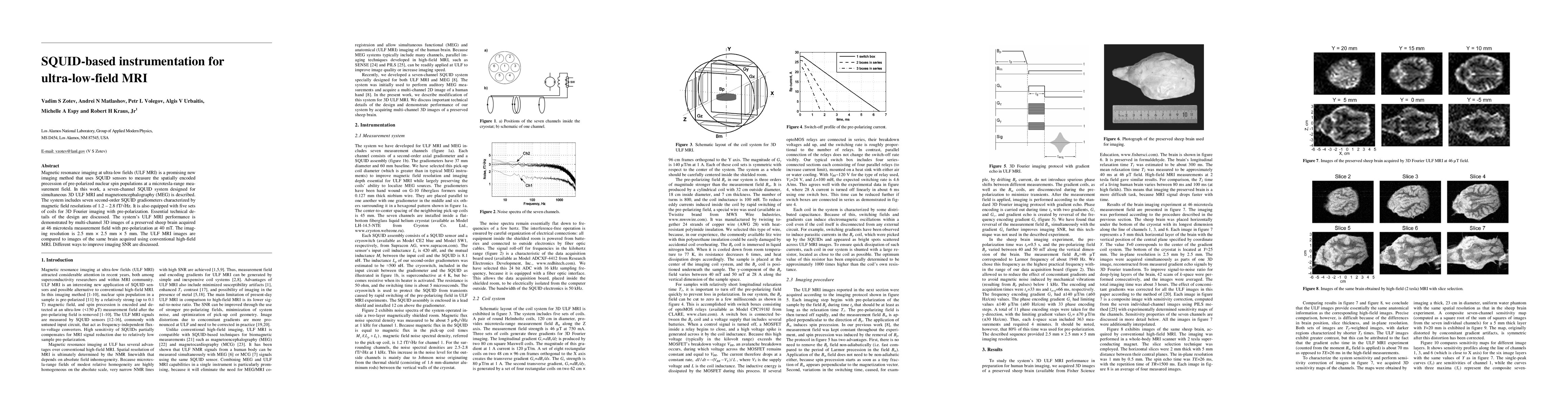
Magnetic resonance imaging at ultra-low fields (ULF MRI) is a promising new imaging method that uses SQUID sensors to measure the spatially encoded precession of pre-polarized nuclear spin populations at a microtesla-range measurement field. In this work, a seven-channel SQUID system designed for simultaneous 3D ULF MRI and magnetoencephalography (MEG) is described. The system includes seven second-order SQUID gradiometers, characterized by magnetic field resolutions of 1.2 - 2.8 fT/rtHz. It is also equipped with five sets of coils for 3D Fourier imaging with pre-polarization. Essential technical details of the design are discussed. The system's ULF MRI performance is demonstrated by multi-channel 3D images of a preserved sheep brain acquired at 46 microtesla measurement field with pre-polarization at 40 mT. The imaging resolution is 2.5 mm x 2.5 mm x 5 mm. The ULF MRI images are compared to images of the same brain acquired using conventional high-field MRI. Different ways to improve imaging SNR are discussed.
AI Key Findings
Get AI-generated insights about this paper's methodology, results, significance, and more — seven facets brought into focus.
Impact
Paper Details
PDF Preview
Key Terms
Citation Network
Current paper (gray), citations (green), references (blue)
Display is limited for performance on very large graphs.
Discussion 0