This paper proposes two generative adversarial network (GAN) models, SSIM-GAN and DSCSI-GAN, to address color variations in histopathology images, ensuring the preservation of structural and texture information. The DSCSI-GAN model notably improves stain normalization, leading to more consistent and informative images, as demonstrated by superior performance in experiments.
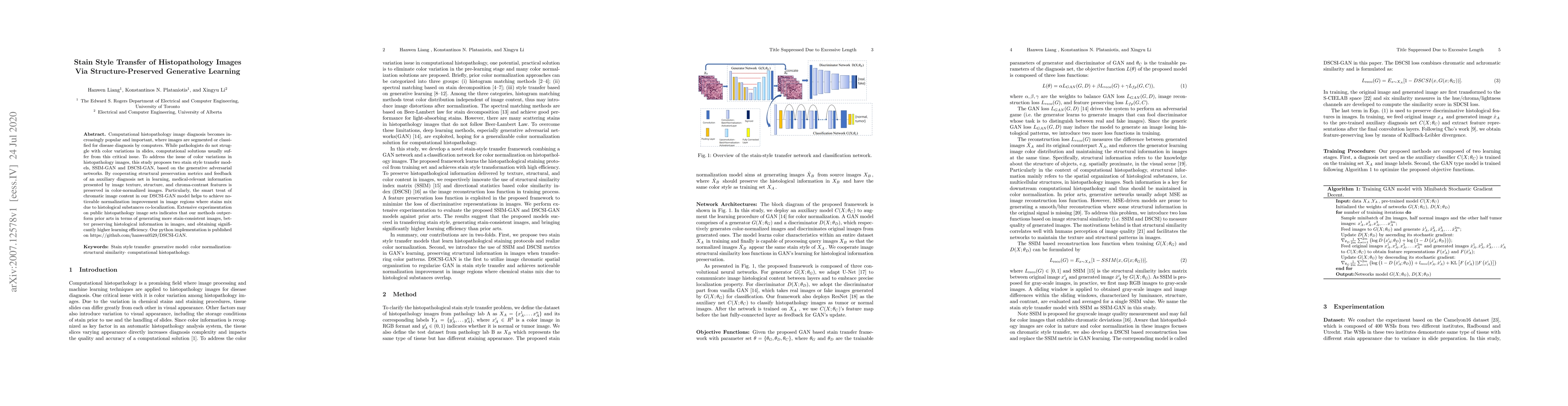
Computational histopathology image diagnosis becomes increasingly popular and important, where images are segmented or classified for disease diagnosis by computers. While pathologists do not struggle with color variations in slides, computational solutions usually suffer from this critical issue. To address the issue of color variations in histopathology images, this study proposes two stain style transfer models, SSIM-GAN and DSCSI-GAN, based on the generative adversarial networks. By cooperating structural preservation metrics and feedback of an auxiliary diagnosis net in learning, medical-relevant information presented by image texture, structure, and chroma-contrast features is preserved in color-normalized images. Particularly, the smart treat of chromatic image content in our DSCSI-GAN model helps to achieve noticeable normalization improvement in image regions where stains mix due to histological substances co-localization. Extensive experimentation on public histopathology image sets indicates that our methods outperform prior arts in terms of generating more stain-consistent images, better preserving histological information in images, and obtaining significantly higher learning efficiency. Our python implementation is published on https://github.com/hanwen0529/DSCSI-GAN.
This graph shows how this paper is connected to other research. The current paper is shown in dark gray, papers that cite it are in green, and papers it references are in blue. You can:
• Hover over any node to see the paper title
• Click a node to view that paper's details
• Drag nodes to rearrange the graph
• Use the zoom controls (bottom right) to zoom in/out
Title | Authors | Year | Actions |
---|---|---|---|
Semi-supervised ViT knowledge distillation network with style transfer normalization for colorectal liver metastases survival prediction
|
Mohamed El Amine Elforaici, E. Montagnon, F. P. Romero, W. Le, F. Azzi, Dominique Trudel, Bich Nguyen, Simon Turcotte, An Tang, Samuel Kadoury
|
2024
|
View Paper |
Generative models for color normalization in digital pathology and dermatology: Advancing the learning paradigm
|
Massimo Salvi, Francesco Branciforti, Filippo Molinari, K. Meiburger
|
2024
|
View Paper |
StainSWIN: Vision transformer-based stain normalization for histopathology image analysis
|
Elif Baykal Kablan, Selen Ayas
|
2024
|
View Paper |
Automated anomaly detection in histology images using deep learning
|
Lillie Shelton, R. Soans, Tosha Shah, Thomas Forest, K. Janardhan, Michael Napolitano, Raymond Gonzalez, Grady Carlson, Jyoti K. Shah, Antong Chen
|
2024
|
View Paper |
Style-Extracting Diffusion Models for Semi-Supervised Histopathology Segmentation
|
Mathias Öttl, Frauke Wilm, Jana Steenpass, Jingna Qiu, M. Rübner, A. Hartmann, Matthias W. Beckmann, Peter A. Fasching, Andreas Maier, R. Erber, Bernhard Kainz, Katharina Breininger
|
2024
|
View Paper |
Semi-supervised ViT knowledge distillation network with style transfer normalization for colorectal liver metastases survival prediction
|
Mohamed El Amine Elforaici, E. Montagnon, F. P. Romero, W. Le, F. Azzi, Dominique Trudel, Bich Nguyen, Simon Turcotte, An Tang, Samuel Kadoury
|
2023
|
View Paper |
Early Prediction of Ductal Carcinoma Histological Subtypes Via Ensemble Transformer Pipelines in Digital Pathology
|
Vrishank Chandrasekhar
|
2023
|
View Paper |
Stain normalization methods for histopathology image analysis: A comprehensive review and experimental comparison
|
Md. Ziaul Hoque, A. Keskinarkaus, Pia Nyberg, T. Seppänen
|
2023
|
View Paper |
Generative Adversarial Networks for Stain Normalisation in Histopathology
|
Jack Breen, K. Zucker, Katie Allen, N. Ravikumar, N. Orsi
|
2023
|
View Paper |
A stain color normalization with robust dictionary learning for breast cancer histological images processing
|
T. A. Tosta, A. D. Freitas, P. D. Faria, L. A. Neves, A. S. Martins, M. Z. Nascimento
|
2023
|
View Paper |
Regional realness-aware generative adversarial networks for stain normalization
|
Elif Baykal Kablan
|
2023
|
View Paper |
A Laplacian Pyramid Based Generative H&E Stain Augmentation Network
|
Fangda Li, Zhiqiang Hu, Wen Chen, A. Kak
|
2023
|
View Paper |
Efficient diagnosis of hematologic malignancies using bone marrow microscopic images: A method based on MultiPathGAN and MobileViTv2
|
Guanghui Yang, Ziqi Qin, Jianmin Mu, H. Mao, Huihui Mao, Min Han
|
2023
|
View Paper |
VSGD-Net: Virtual Staining Guided Melanocyte Detection on Histopathological Images
|
Kechun Liu, Beibin Li, Wenjun Wu, Caitlin J. May, Oliver H. Chang, S. Knezevich, L. Reisch, J. Elmore, Linda G. Shapiro
|
2023
|
View Paper |
Stain normalization in digital pathology: Clinical multi-center evaluation of image quality
|
Nicola Michielli, Alessandro Caputo, Manuela Scotto, Alessandro Mogetta, Orazio Pennisi, F. Molinari, D. Balmativola, M. Bosco, A. Gambella, J. Metovic, Daniele Tota, L. Carpenito, P. Gasparri, M. Salvi
|
2022
|
View Paper |
Style Transfer with Bio-realistic Appearance Manipulation for Skin-tone Inclusive rPPG
|
Yunhao Ba, Zhen Wang, Kerim Doruk Karinca, Oyku Deniz Bozkurt, A. Kadambi
|
2022
|
View Paper |
Built to last? Reproducibility and Reusability of Deep Learning Algorithms in Computational Pathology
|
S. Wagner, C. Matek, Sayedali Shetab Boushehri, M. Boxberg, L. Lamm, A. Sadafi, Dominik Jens Elias Waibel, C. Marr, T. Peng
|
2022
|
View Paper |
RestainNet: a self-supervised digital re-stainer for stain normalization
|
Bingchao Zhao, Jiatai Lin, C. Liang, Zongjian Yi, Xin Chen, Bingbing Li, Weihao Qiu, Dan Li, Li Liang, Chu Han, Zaiyi Liu
|
2022
|
View Paper |
Structure-Preserving Multi-Domain Stain Color Augmentation using Style-Transfer with Disentangled Representations
|
S. Wagner, Nadieh Khalili, Raghav Sharma, M. Boxberg, C. Marr, Walter de Back, Tingying Peng
|
2021
|
View Paper |
Smart-phone phase contrast microscope with a singlet lens and deep learning
|
Yinxu Bian, Yannan Jiang, Yuran Huang, Xiaofei Yang, Weijie Deng, Huaxia Shen, Renbin Shen, C. Kuang
|
2021
|
View Paper |
Evaluating Registrations of Serial Sections With Distortions of the Ground Truths
|
Oleg Lobachev, Takuya Funatomi, Alexander Pfaffenroth, R. Förster, Lars Knudsen, C. Wrede, M. Guthe, D. Haberthür, R. Hlushchuk, T. Salaets, J. Toelen, Simone Gaffling, C. Mühlfeld, R. Grothausmann
|
2020
|
View Paper |
Deep learning colorful ptychographic iterative engine lens-less diffraction microscopy
|
Yinxu Bian, Yannan Jiang, Jiaxiong Wang, Shen Yang, Weijie Deng, Xiaofei Yang, Renbin Shen, Huaxia Shen, C. Kuang
|
2022
|
View Paper |
Whole Slide Image Quality in Digital Pathology: Review and Perspectives
|
Romain Brixtel, Sébastien Bougleux, O. Lézoray, Yann Caillot, Benoît Lemoine, Mathieu Fontaine, Dalal Nebati, Arnaud Renouf
|
2022
|
View Paper |
Published: 2021-06-04
Igor Zingman, Fabian Heinemann, Sergio Frayle, Ivan Tankoyeu, Segrey Sukhanov
Published: 2023-04-07
Published: 2020-10-07
Comments (0)