Authors
Summary
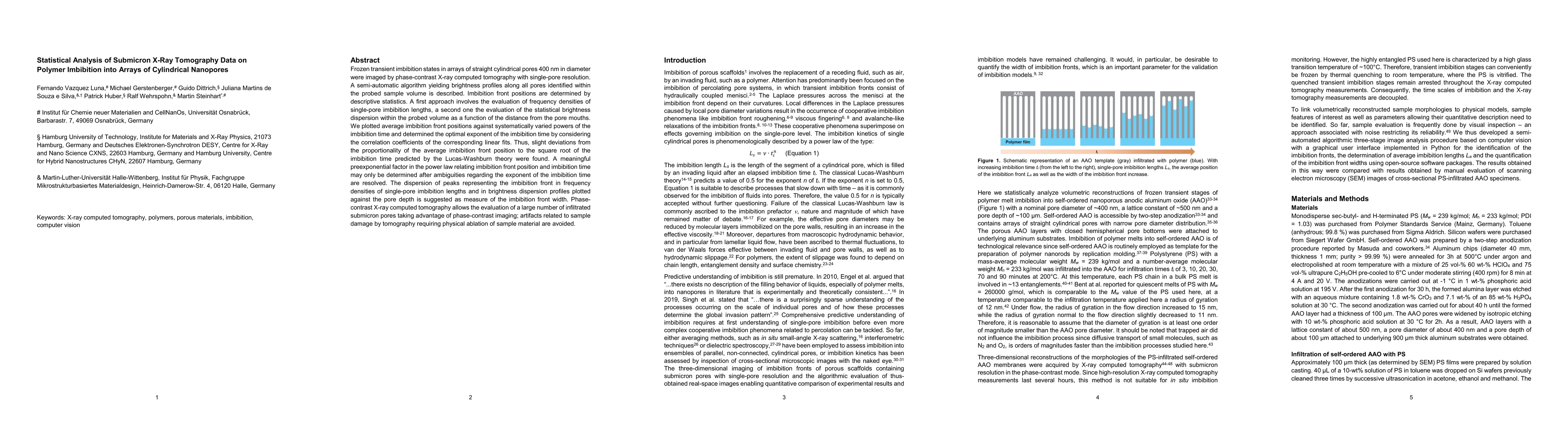
Frozen transient imbibition states in arrays of straight cylindrical pores 400 nm in diameter were imaged by phase-contrast X-ray computed tomography with single-pore resolution. A semi-automatic algorithm yielding brightness profiles along all pores identified within the probed sample volume is described. Imbibition front positions are determined by descriptive statistics. A first approach involves the evaluation of frequency densities of single-pore imbibition lengths, a second one the evaluation of the statistical brightness dispersion within the probed volume as a function of the distance from the pore mouths. We plotted average imbibition front positions against systematically varied powers of the imbibition time and determined the optimal exponent of the imbibition time by considering the correlation coefficients of the corresponding linear fits. Thus, slight deviations from the proportionality of the average imbibition front position to the square root of the imbibition time predicted by the Lucas-Washburn theory were found. A meaningful preexponential factor in the power law relating imbibition front position and imbibition time may only be determined after ambiguities regarding the exponent of the imbibition time are resolved. The dispersion of peaks representing the imbibition front in frequency densities of single-pore imbibition lengths and in brightness dispersion profiles plotted against the pore depth is suggested as measure of the imbibition front width. Phase-contrast X-ray computed tomography allows the evaluation of a large number of infiltrated submicron pores taking advantage of phase-contrast imaging; artifacts related to sample damage by tomography requiring physical ablation of sample material are avoided.
AI Key Findings
Get AI-generated insights about this paper's methodology, results, and significance.
Paper Details
PDF Preview
Key Terms
Citation Network
Current paper (gray), citations (green), references (blue)
Display is limited for performance on very large graphs.
Similar Papers
Found 4 papersStraight versus Spongy -- Effect of Tortuosity on Polymer Imbibition into Nanoporous Matrices Assessed by Segmentation-Free Analysis of 3D Sample Reconstructions
Patrick Huber, Martin Steinhart, Dirk Enke et al.
Imbibition of Oil in Dry and Prewetted Calcite Nanopores
Rui Qiao, Shihao Wang, Ejaz Ahmed et al.
Deformation Dynamics of Nanopores upon Water Imbibition
Howard A. Stone, Patrick Huber, Robert Meissner et al.
| Title | Authors | Year | Actions |
|---|
Comments (0)