Statistical analysis of virion-cell interactions mediated by peptide nanofibrils and peptide amphiphiles using STEM tomography
Publication
Metrics
Paper Preview
Abstract
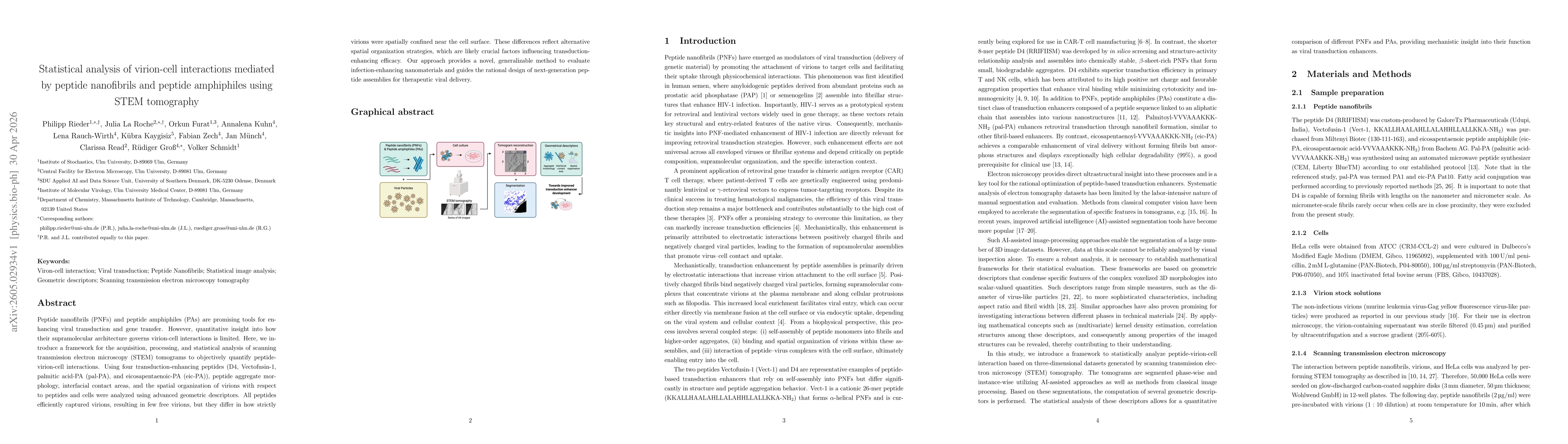
Peptide nanofibrils (PNFs) and peptide amphiphiles (PAs) are promising tools for enhancing viral transduction and gene transfer. However, quantitative insight into how their supramolecular architecture governs virion-cell interactions is limited. Here, we introduce a framework for the acquisition, processing, and statistical analysis of scanning transmission electron microscopy (STEM) tomograms to objectively quantify peptide-virion-cell interactions. Using four transduction-enhancing peptides (D4, Vectofusin-1, palmitic acid-PA (pal-PA), and eicosapentaenoic-PA (eic-PA)), peptide aggregate morphology, interfacial contact areas, and the spatial organization of virions with respect to peptides and cells were analyzed using advanced geometric descriptors. All peptides efficiently captured virions, resulting in few free virions, but they differ in how strictly virions were spatially confined near the cell surface. These differences reflect alternative spatial organization strategies, which are likely crucial factors influencing transduction-enhancing efficacy. Our approach provides a novel, generalizable method to evaluate infection-enhancing nanomaterials and guides the rational design of next-generation peptide assemblies for therapeutic viral delivery.
AI Key Findings
Get AI-generated insights about this paper's methodology, results, significance, and more — seven facets brought into focus.
Discussion 0