Summary
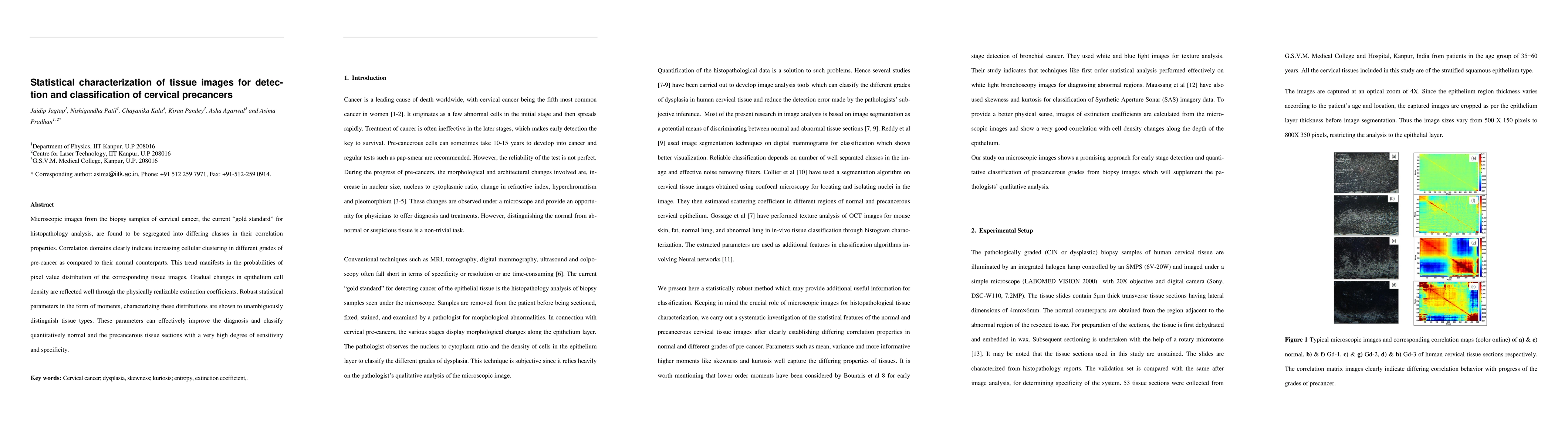
Microscopic images from the biopsy samples of cervical cancer, the current "gold standard" for histopathology analysis, are found to be segregated into differing classes in their correlation properties. Correlation domains clearly indicate increasing cellular clustering in different grades of pre-cancer as compared to their normal counterparts. This trend manifests in the probabilities of pixel value distribution of the corresponding tissue images. Gradual changes in epithelium cell density are reflected well through the physically realizable extinction coefficients. Robust statistical parameters in the form of moments, characterizing these distributions are shown to unambiguously distinguish tissue types. These parameters can effectively improve the diagnosis and classify quantitatively normal and the precancerous tissue sections with a very high degree of sensitivity and specificity.
AI Key Findings
Get AI-generated insights about this paper's methodology, results, and significance.
Paper Details
PDF Preview
Key Terms
Citation Network
Current paper (gray), citations (green), references (blue)
Display is limited for performance on very large graphs.
| Title | Authors | Year | Actions |
|---|
Comments (0)