ArxivLens

Publication

Published: Aug 11, 2024
Categories: q-bio.NC

Metrics

Source: ArXiv

AI Quick Summary

This study found that individuals with temporal lobe epilepsy and a history of status epilepticus exhibited significant thinning of the entorhinal cortex, particularly on the ipsilateral side, compared to those without a history of status epilepticus. These results suggest the entorhinal cortex may be particularly vulnerable to prolonged seizures.

Quick Answers

What is "Status epilepticus and thinning of the entorhinal cortex" about?

This study found that individuals with temporal lobe epilepsy and a history of status epilepticus exhibited significant thinning of the entorhinal cortex, particularly on the ipsilateral side, compared to those without a history of status epilepticus. These results suggest the entorhinal cortex may be particularly vulnerable to prolonged seizures.

What methodology did the authors use?

The study used a large cohort of individuals with TLE, exploring the association between reduced cortical thickness in the ipsilateral and contralateral entorhinal cortex and a history of SE. More in Methodology →

What are the key results?

Reduced cortical thickness in the ipsilateral entorhinal cortex was found to be associated with a history of SE — Increased subcortical volume of the ipsilateral hippocampus was observed in individuals with a history of SE More in Key Results →

Why is this work significant?

The study highlights the potential impact of SE on brain structure and function, shedding light on the underlying mechanisms of epilepsy. More in Significance →

What are the main limitations?

Potential bias in patient selection due to presurgical evaluation — Age range limited to adults only More in Limitations →

Paper Preview

Abstract

Status epilepticus (SE) carries risks of morbidity and mortality. Experimental studies have implicated the entorhinal cortex in prolonged seizures; however, studies in large human cohorts are limited. We hypothesised that individuals with temporal lobe epilepsy (TLE) and a history of SE would have more severe entorhinal atrophy compared to others with TLE and no history of SE. 357 individuals with drug resistant temporal lobe epilepsy (TLE) and 100 healthy controls were scanned on a 3T MRI. For all subjects the cortex was segmented, parcellated, and the thickness calculated from the T1-weighted anatomical scan. Subcortical volumes were derived similarly. Cohen's d and Wilcoxon rank-sum tests respectively were used to capture effect sizes and significance. 
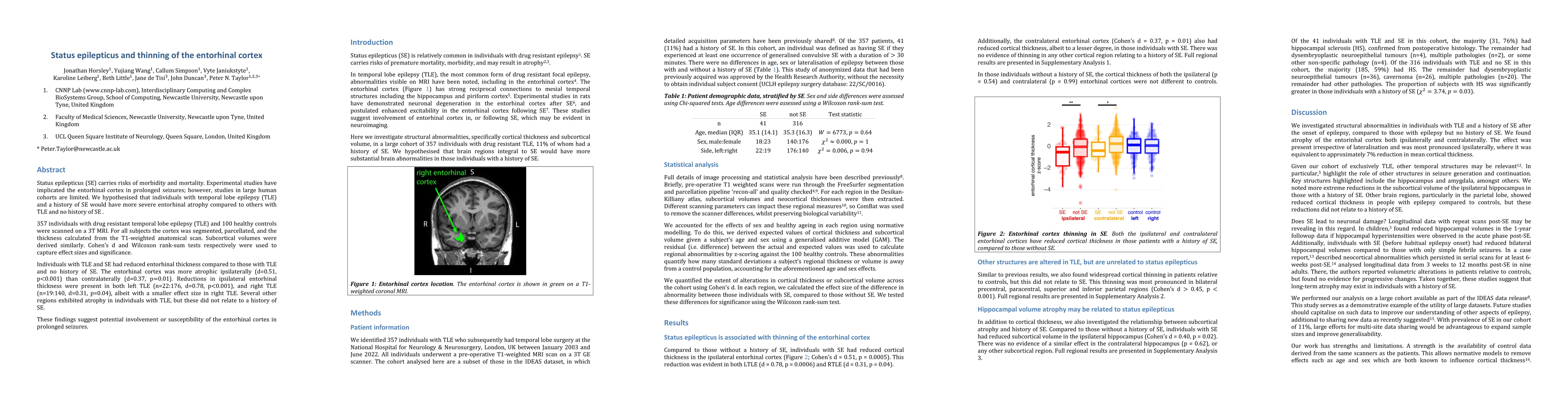
Individuals with TLE and SE had reduced entorhinal thickness compared to those with TLE and no history of SE. The entorhinal cortex was more atrophic ipsilaterally (d=0.51, p<0.001) than contralaterally (d=0.37, p=0.01). Reductions in ipsilateral entorhinal thickness were present in both left TLE (n=22:176, d=0.78, p<0.001), and right TLE (n=19:140, d=0.31, p=0.04), albeit with a smaller effect size in right TLE. Several other regions exhibited atrophy in individuals with TLE, but these did not relate to a history of SE. These findings suggest potential involvement or susceptibility of the entorhinal cortex in prolonged seizures.

Key Findings, in focus

Seven facets of this paper, analysed and brought into focus by AI.

AI-generated · verify against source Generated Sep 06, 2025
In focus · one-line synthesis

The study highlights the potential impact of SE on brain structure and function, shedding light on the underlying mechanisms of epilepsy.

01
MethodologyHow they did it

The study used a large cohort of individuals with TLE, exploring the association between reduced cortical thickness in the ipsilateral and contralateral entorhinal cortex and a history of SE.

02
ResultsWhat they found
  • Reduced cortical thickness in the ipsilateral entorhinal cortex was found to be associated with a history of SE
  • Increased subcortical volume of the ipsilateral hippocampus was observed in individuals with a history of SE
  • Other brain regions, particularly in the parietal lobe, showed reduced cortical thickness in people with epilepsy compared to controls
03
SignificanceWhy it matters

The study highlights the potential impact of SE on brain structure and function, shedding light on the underlying mechanisms of epilepsy.

04
Technical ContributionsWhat's new technically

The study leverages large-scale imaging data to identify novel associations between SE and brain anatomy.

05
NoveltyWhat sets it apart

This work contributes to the understanding of SE's impact on brain structure, highlighting its potential as a risk factor for epilepsy

06
LimitationsWhat to be cautious about
  • Potential bias in patient selection due to presurgical evaluation
  • Age range limited to adults only
07
Future WorkWhere it goes next
  • Investigating the frequency and duration of SE to better understand its effects on brain structure
  • Exploring other temporal structures relevant to seizure generation and continuation

Impact

16
References

Paper Details

Paper ID: 2408.05789
Categories:
q-bio.NC

How to Cite This Paper

Reference manager: RIS EndNote

Citation Network

Current paper (gray), citations (green), references (blue)

Display is limited for performance on very large graphs.

Current Paper
Citations
References
Click to view

Related Papers

4 found


Discussion 0

This website uses cookies to ensure you get the best experience. Learn more