Quick Summary:

This paper proposes training strategies for developing stain invariant convolutional neural networks (CNNs) to analyze digitized whole slide images from differently stained tissue sections in digital pathology. The unsupervised methods presented show significant improvements over standard training strategies by enabling the network to generalize across various staining modalities with minimal additional annotations.

Summary

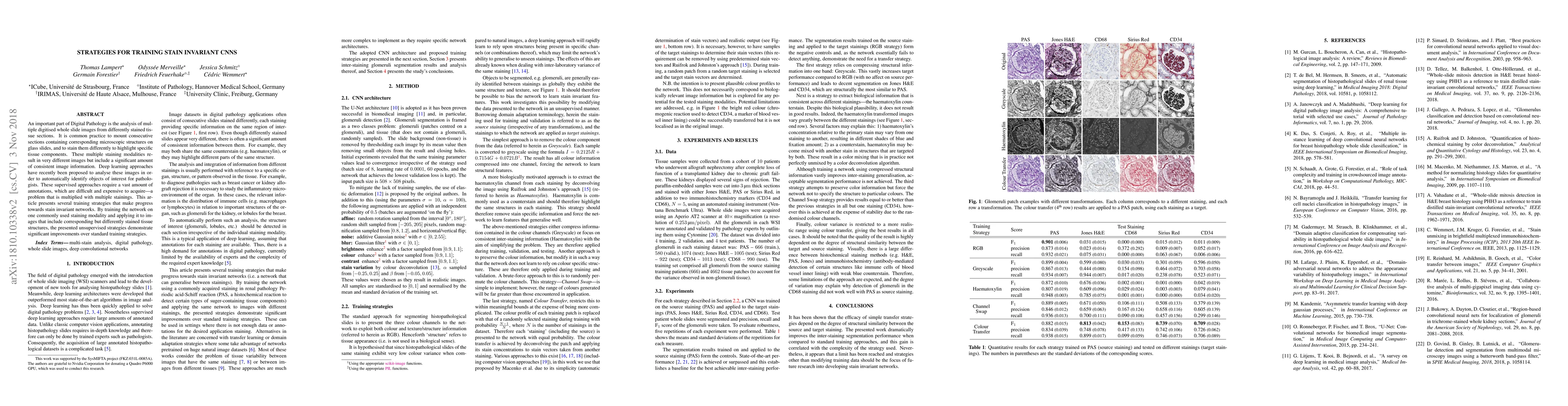
An important part of Digital Pathology is the analysis of multiple digitised whole slide images from differently stained tissue sections. It is common practice to mount consecutive sections containing corresponding microscopic structures on glass slides, and to stain them differently to highlight specific tissue components. These multiple staining modalities result in very different images but include a significant amount of consistent image information. Deep learning approaches have recently been proposed to analyse these images in order to automatically identify objects of interest for pathologists. These supervised approaches require a vast amount of annotations, which are difficult and expensive to acquire---a problem that is multiplied with multiple stainings. This article presents several training strategies that make progress towards stain invariant networks. By training the network on one commonly used staining modality and applying it to images that include corresponding but differently stained tissue structures, the presented unsupervised strategies demonstrate significant improvements over standard training strategies.

AI Key Findings

Get AI-generated insights about this paper's methodology, results, and significance.

Paper Details

PDF Preview

Download PDF

Key Terms

differently (0.301)
strategies (0.228)
images (0.228)
sections (0.208)
training (0.189)
multiple (0.180)
include (0.178)
structures (0.157)
annotations (0.149)
approaches (0.148)
modality (0.147)
corresponding (0.144)
significant (0.144)
vast (0.142)
modalities (0.137)
glass (0.131)
analyse (0.130)
commonly used (0.130)
recently proposed (0.129)
unsupervised (0.129)

Citation Network

Current paper (gray), citations (green), references (blue)

Current Paper
Citations
References
Click to view
Title Authors Year Actions
Maximising Histopathology Segmentation using Minimal Labels via Self-Supervision
Zeeshan Nisar, Thomas Lampert
2024
View Paper
Unsupervised Latent Stain Adaptation for Computational Pathology
Daniel Reisenbüchler, Lucas Luttner, N. Schaadt, Friedrich Feuerhake, D. Merhof
2024
View Paper
Unsupervised Multi-Domain Progressive Stain Transfer Guided by Style Encoding Dictionary
Xianchao Guan, Yifeng Wang, Yi‐Ting Lin, Xi Li, Yongbing Zhang
2024
View Paper
Unpaired virtual histological staining using prior-guided generative adversarial networks
Renao Yan, Qiming He, Yiqing Liu, Peng Ye, Liang Zhu, Shanshan Shi, Jizhou Gou, Yonghong He, Tian Guan, Guangde Zhou
2023
View Paper
HistoStarGAN: A Unified Approach to Stain Normalisation, Stain Transfer and Stain Invariant Segmentation in Renal Histopathology
Jelica Vasiljevi'c, F. Feuerhake, Cédric Wemmert, T. Lampert
2022
View Paper
Conditional image synthesis for improved segmentation of glomeruli in renal histopathological images
Florian Allender, Rémi Allègre, Cédric Wemmert, J. Dischler
2022
View Paper
CS-CO: A Hybrid Self-Supervised Visual Representation Learning Method for H&E-stained Histopathological Images
Pengshuai Yang, Xiaoxu Yin, Haiming Lu, Zhongliang Hu, Xuegong Zhang, Rui Jiang, Hairong Lv
2022
View Paper
Data augmentation based on spatial deformations for histopathology: An evaluation in the context of glomeruli segmentation
Florian Allender, Rémi Allègre, Cédric Wemmert, J. Dischler
2022
View Paper
Towards Measuring Domain Shift in Histopathological Stain Translation in an Unsupervised Manner
Zeeshan Nisar, Jelica Vasiljević, P. Gançarski, T. Lampert
2022
View Paper
Improving unsupervised stain-to-stain translation using self-supervision and meta-learning
Nassim Bouteldja, B. Klinkhammer, Tarek Schlaich, P. Boor, D. Merhof
2021
View Paper
An automatic framework for fusing information from differently stained consecutive digital whole slide images: A case study in renal histology
Odyssée Merveille, T. Lampert, J. Schmitz, G. Forestier, F. Feuerhake, Cédric Wemmert
2021
View Paper
Self Adversarial Attack as an Augmentation Method for Immunohistochemical Stainings
Jelica Vasiljevi'c, F. Feuerhake, Cédric Wemmert, T. Lampert
2021
View Paper
Inter-vendor performance of deep learning in segmenting acute ischemic lesions on diffusion-weighted imaging: a multicenter study
D. Alis, M. Yergin, Ceren Aliş, Ç. Topel, O. Asmakutlu, Omer Bagcilar, Y. Senli, Ahmet Ustundag, Vefa Salt, S. N. Dogan, M. Velioglu, H. Selçuk, B. Kara, I. Oksuz, O. Kızılkılıç, E. Karaarslan
2020
View Paper
Towards Histopathological Stain Invariance by Unsupervised Domain Augmentation using Generative Adversarial Networks
Jelica Vasiljevi'c, F. Feuerhake, Cédric Wemmert, T. Lampert
2020
View Paper
An automatic framework to study the tissue micro-environment of renal glomeruli in differently stained consecutive digital whole slide images
Odyssée Merveille, T. Lampert, J. Schmitz, G. Forestier, F. Feuerhake, Cédric Wemmert
2020
View Paper
Advancing Medical Imaging Informatics by Deep Learning-Based Domain Adaptation
A. Choudhary, L. Tong, Yuanda Zhu, May D. Wang
2020
View Paper
Real-Time Detection of Glomeruli in Renal Pathology
Robin Heckenauer, J. Weber, Cédric Wemmert, F. Feuerhake, M. Hassenforder, Pierre-Alain Muller, G. Forestier
2020
View Paper
Towards Staining Independent Segmentation of Glomerulus from Histopathological Images of Kidney
Robin Liu, Lu Wang, Jim He, Wenfang Chen
2019
View Paper
Unsupervised Latent Stain Adaption for Digital Pathology
Daniel Reisenbüchler, Lucas Luttner, N. Schaadt, Friedrich Feuerhake, D. Merhof
2024
View Paper
Deep Learning for Histopathological Image Analysis
Cédric Wemmert, J. Weber, F. Feuerhake, G. Forestier
2021
View Paper

Comments (0)

This website uses cookies to ensure you get the best experience. Learn more