Quick Answers
What methodology did the authors use?
The research introduces XMAG, a 5x magnification foundation model for computational pathology, utilizing cross-magnification distillation and multiple instance learning (MIL) for whole-slide image analysis. It evaluates performance on various cancer datasets using linear probing and compares against 20x models. More in Methodology →
What are the key results?
XMAG achieves comparable accuracy (0.878) to 20x UNI2 on SPIDER-Breast dataset but lower AUC (0.988 vs 0.994) — On SPIDER-CRC, XMAG shows slightly lower performance (AUC 0.990) compared to 20x models (0.994) More in Key Results →
Why is this work significant?
This research advances computational pathology by enabling efficient, lower-magnification analysis for histopathological tasks, potentially reducing computational demands without sacrificing diagnostic accuracy. More in Significance →
What are the main limitations?
Performance slightly lags behind 20x models in some tasks — Limited to specific cancer types and datasets evaluated More in Limitations →
Abstract
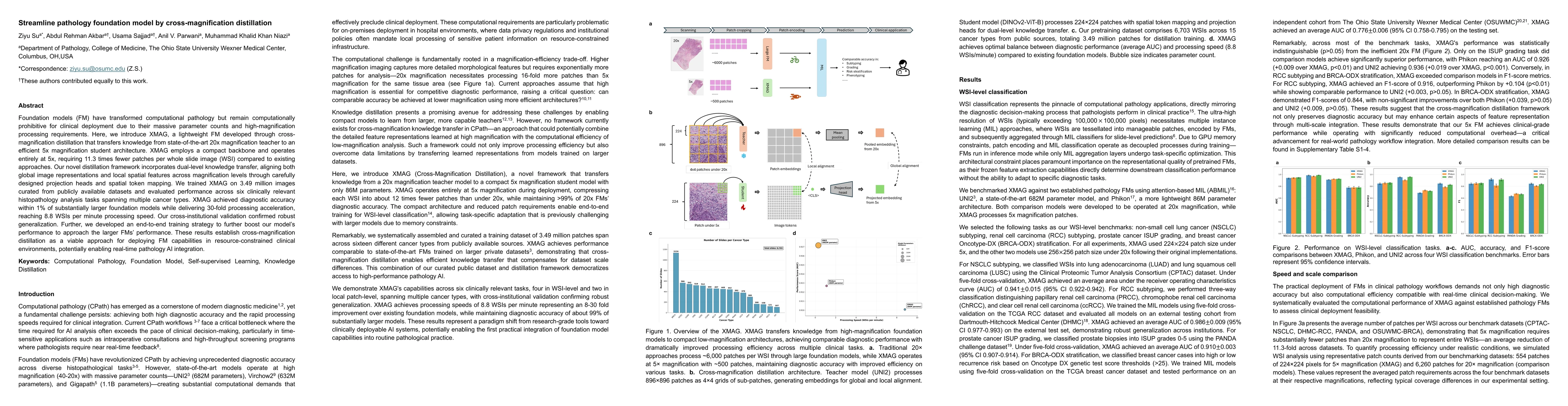
Foundation models (FM) have transformed computational pathology but remain computationally prohibitive for clinical deployment due to their massive parameter counts and high-magnification processing requirements. Here, we introduce XMAG, a lightweight FM developed through corss-magnification distillation that transfers knowledge from state-of-the-art 20x magnification teacher to an efficient 5x magnification student architecture. XMAG employs a compact backbone and operates entirely at 5x, requiring 11.3 times fewer patches per whole slide image (WSI) compared to existing approaches. Our Novel distillation framework incorporates dual-level knowledge transfer, aligning both global image representations and local spatial token mapping. We trained XMAG on 3.49 million images curated from publicly available datasets and evaluated performance across six clinically relevant histopathology analysis tasks spanning multiple cancer types. XMAG achieved diagnostic accuracy within 1% of substantially larger foundation models while delivering 30-fold processing acceleration, reaching 8.8 WSIs per minute processing speed. Our cross-institutional validation confirmed robust generalization. Further, we developed an end-to-end training strategy to further boost our model's performance to approach the larger FMs' performance. These results establish cross-magnification distillation as a viable approach for deploying FM capabilities in resource-constrained clinical environments, potentially enabling real-time pathology AI integration.
AI Key Findings
Generated Oct 01, 2025
Methodology — What approach did the authors take?
The research introduces XMAG, a 5x magnification foundation model for computational pathology, utilizing cross-magnification distillation and multiple instance learning (MIL) for whole-slide image analysis. It evaluates performance on various cancer datasets using linear probing and compares against 20x models.
Key Results — What are the main findings?
- XMAG achieves comparable accuracy (0.878) to 20x UNI2 on SPIDER-Breast dataset but lower AUC (0.988 vs 0.994)
- On SPIDER-CRC, XMAG shows slightly lower performance (AUC 0.990) compared to 20x models (0.994)
- XMAG demonstrates efficient computation with 86M parameters vs 682M in 20x models while maintaining competitive performance
Significance — Why does this research matter?
This research advances computational pathology by enabling efficient, lower-magnification analysis for histopathological tasks, potentially reducing computational demands without sacrificing diagnostic accuracy.
Technical Contribution — What is the technical contribution?
Proposes cross-magnification distillation framework for training efficient foundation models in computational pathology, combined with multiple instance learning for whole-slide image analysis.
Novelty — What is new about this work?
First application of cross-magnification distillation in computational pathology, enabling effective analysis at lower magnification levels while maintaining diagnostic performance comparable to high-magnification models.
Limitations — What are the limitations of this study?
- Performance slightly lags behind 20x models in some tasks
- Limited to specific cancer types and datasets evaluated
Future Work — What did the authors propose for future work?
- Explore application across more cancer types and histopathological tasks
- Investigate hybrid approaches combining different magnification levels
- Enhance model robustness for varied histopathological patterns
How to Cite This Paper
@article{su2025streamline,
title = {Streamline pathology foundation model by cross-magnification
distillation},
author = {Su, Ziyu and Akbar, Abdul Rehman and Sajjad, Usama and others},
year = {2025},
eprint = {2509.23097},
archivePrefix = {arXiv},
primaryClass = {cs.CV},
}
Su, Z., Akbar, A., Sajjad, U., Parwani, A., & Niazi, M. (2025). Streamline pathology foundation model by cross-magnification
distillation. arXiv. https://arxiv.org/abs/2509.23097
Su, Ziyu, et al. "Streamline pathology foundation model by cross-magnification
distillation." arXiv, 2025, arxiv.org/abs/2509.23097.
PDF Preview
Similar Papers
Found 4 papersTowards A Generalizable Pathology Foundation Model via Unified Knowledge Distillation
Jiabo Ma, Yihui Wang, Yingxue Xu et al.
Mind the Gap: Continuous Magnification Sampling for Pathology Foundation Models
Alexander Möllers, Julius Hense, Florian Schulz et al.
A Novel Pathology Foundation Model by Mayo Clinic, Charité, and Aignostics
Maximilian Alber, Stephan Tietz, Jonas Dippel et al.
Comments (0)