Summary
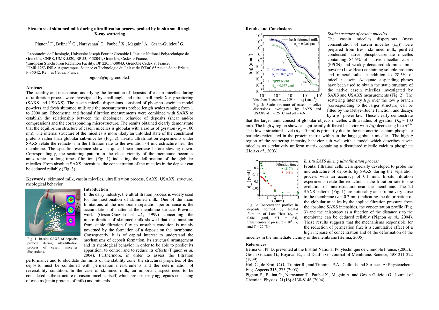
The stability and mechanism underlying the formation of deposits of casein micelles during ultrafiltration process were investigated by small-angle (SAXS) and ultra small-angle X-ray scattering(USAXS) which allowed us to probe the structure of skimmed milk on an exceptionally wide range of length scales from 1 to 2000 nm. Frontal filtration cells were specially developed to probe the microstructure of deposits by SAXS during the separation process. The results revealed two characteristic length scales for the equilibrium structure with radius of gyrations Rg, about 100 nm and 5.6 nm, pertaining to the globular micelles and their non-globular internal structure respectively (Pignon et al. 2004). In-situ scattering measurements showed that the decrease of permeation flows is directly related to the deformation and compression of the micelles in the immediate vicinity of the membrane (figure 1). From absolute SAXS intensities, the concentration of the micelles in the deposit can be deduced reliably.
AI Key Findings
Get AI-generated insights about this paper's methodology, results, and significance.
Paper Details
PDF Preview
Key Terms
Similar Papers
Found 4 papersIn situ small-angle X-ray scattering reveals strong condensation of DNA origami during silicification
Bert Nickel, Martina F. Ober, Anna Baptist et al.
No citations found for this paper.

Comments (0)