Structure-preserving Guided Retinal Image Filtering and Its Application for Optic Disc Analysis
Publication
Metrics
AI Quick Summary
This paper proposes a novel structure-preserving guided retinal image filtering (SGRIF) method to restore the quality of retinal images degraded by human-lens attenuation and scattering. The SGRIF method improves image contrast and enhances subsequent optic disc analysis tasks, including deep learning-based optic cup segmentation and sparse learning-based cup-to-disc ratio computation.
Paper Preview
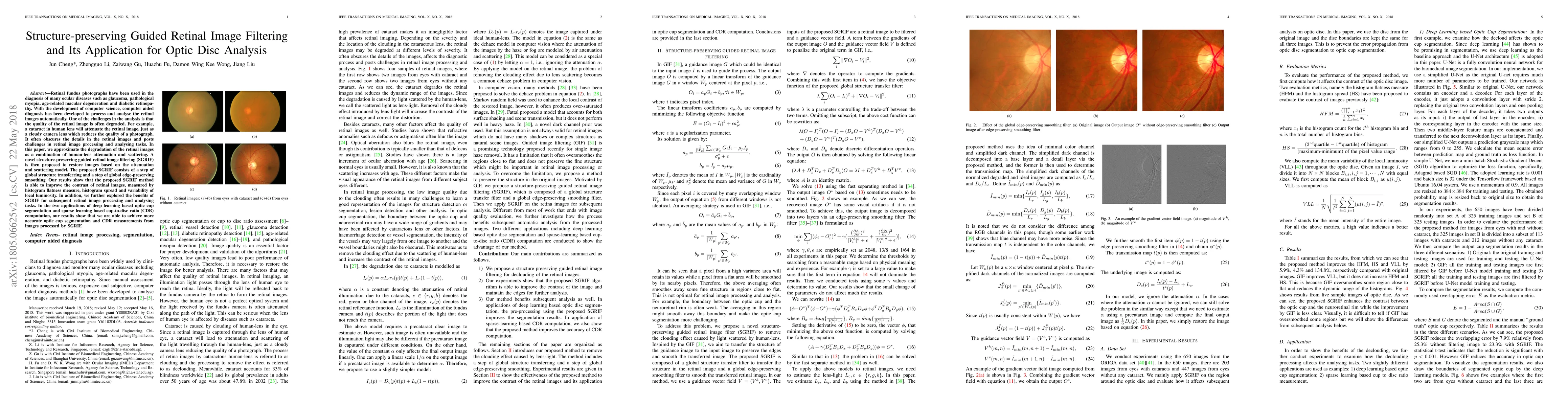
Abstract
Retinal fundus photographs have been used in the diagnosis of many ocular diseases such as glaucoma, pathological myopia, age-related macular degeneration and diabetic retinopathy. With the development of computer science, computer aided diagnosis has been developed to process and analyse the retinal images automatically. One of the challenges in the analysis is that the quality of the retinal image is often degraded. For example, a cataract in human lens will attenuate the retinal image, just as a cloudy camera lens which reduces the quality of a photograph. It often obscures the details in the retinal images and posts challenges in retinal image processing and analysing tasks. In this paper, we approximate the degradation of the retinal images as a combination of human-lens attenuation and scattering. A novel structure-preserving guided retinal image filtering (SGRIF) is then proposed to restore images based on the attenuation and scattering model. The proposed SGRIF consists of a step of global structure transferring and a step of global edge-preserving smoothing. Our results show that the proposed SGRIF method is able to improve the contrast of retinal images, measured by histogram flatness measure, histogram spread and variability of local luminosity. In addition, we further explored the benefits of SGRIF for subsequent retinal image processing and analysing tasks. In the two applications of deep learning based optic cup segmentation and sparse learning based cup-to-disc ratio (CDR) computation, our results show that we are able to achieve more accurate optic cup segmentation and CDR measurements from images processed by SGRIF.
AI Key Findings
Get AI-generated insights about this paper's methodology, results, significance, and more — seven facets brought into focus.
Impact
Paper Details
PDF Preview
Key Terms
Citation Network
Current paper (gray), citations (green), references (blue)
Display is limited for performance on very large graphs.
Discussion 0