Study of SARS-CoV-2 Spike Protein by Surface Enhanced Raman Spectroscopy and Transmission Electron Microscopy
Publication
Metrics
AI Quick Summary
This study uses transmission electron microscopy (TEM) and surface enhanced Raman spectroscopy (SERS) to analyze the SARS-CoV-2 spike protein's structure, identifying characteristic Raman shifts and periodic lattice orientations, which could inform the development of SERS-based diagnostic tools for COVID-19.
Paper Preview
Abstract
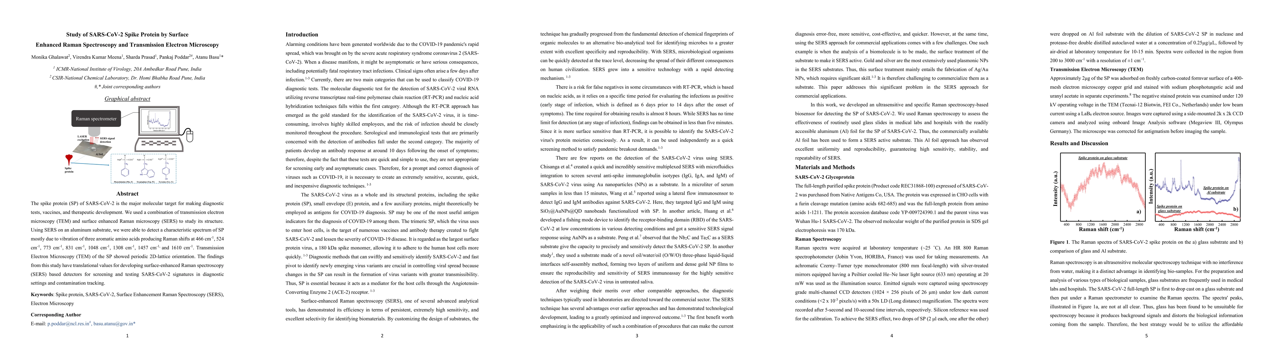
The spike protein (SP) of SARS-CoV-2 is the major molecular target for making diagnostic tests, vaccines, and therapeutic development. We used a combination of transmission electron microscopy (TEM) and surface enhanced Raman microscopy (SERS) to study its structure. Using SERS on an aluminum substrate, we were able to detect a characteristic spectrum of SP mostly due to vibration of three aromatic amino acids producing Raman shifts at 466 cm-1, 524 cm-1, 773 cm-1, 831 cm-1, 1048 cm-1, 1308 cm-1, 1457 cm-1, and 1610 cm-1. Transmission Electron Microscopy (TEM) of the SP showed periodic 2D-lattice orientation. The findings from this study have translational values for developing surface-enhanced Raman spectroscopy (SERS) based detectors for screening and testing SARS-CoV-2 signatures in diagnostic settings and contamination tracking.
AI Key Findings
Get AI-generated insights about this paper's methodology, results, significance, and more — seven facets brought into focus.
Paper Details
Authors
PDF Preview
Related Papers
No references found for this paper.
Discussion 0