Sub-nanometer-thick native sp2 carbon on oxidized diamond surfaces
Publication
Metrics
AI Quick Summary
This research utilizes angle-resolved XPS to analyze the first ten nanometers of oxygen-terminated diamond, identifying functional groups and quantifying a 0.4 ± 0.1 nm thick sp2 carbon layer on the surface, indicating most oxygen bonds with this layer rather than the bulk sp3 carbon.
Paper Preview
Abstract
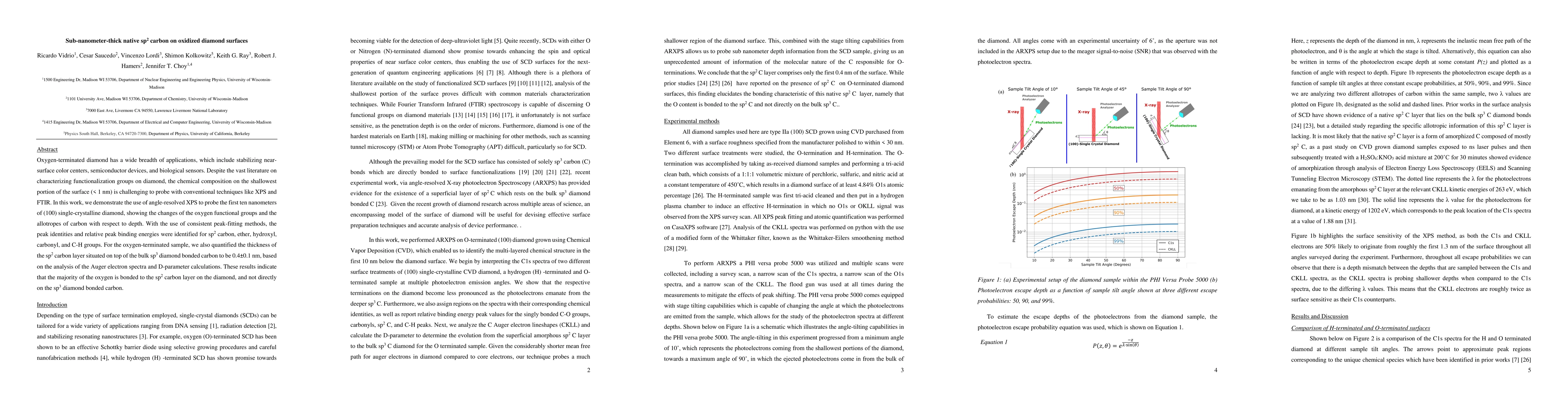
Oxygen-terminated diamond has a wide breadth of applications, which include stabilizing near-surface color centers, semiconductor devices, and biological sensors. Despite the vast literature on characterizing functionalization groups on diamond, the chemical composition on the shallowest portion of the surface (< 1 nm) is challenging to probe with conventional techniques like XPS and FTIR. In this work, we demonstrate the use of angle-resolved XPS to probe the first ten nanometers of (100) single-crystalline diamond, showing the changes of the oxygen functional groups and the allotropes of carbon with respect to depth. With the use of consistent peak-fitting methods, the peak identities and relative peak binding energies were identified for sp2 carbon, ether, hydroxyl, carbonyl, and C-H groups. For the oxygen-terminated sample, we also quantified the thickness of the sp2 carbon layer situated on top of the bulk sp3 diamond bonded carbon to be 0.4 $\pm$ 0.1 nm, based on the analysis of the Auger electron spectra and D-parameter calculations. These results indicate that the majority of the oxygen is bonded to the sp2 carbon layer on the diamond, and not directly on the sp3 diamond bonded carbon.
AI Key Findings
Get AI-generated insights about this paper's methodology, results, significance, and more — seven facets brought into focus.
Impact
Authors
PDF Preview
Citation Network
Current paper (gray), citations (green), references (blue)
Display is limited for performance on very large graphs.
Discussion 0