AI Quick Summary
This study develops an MRI fusion technique to classify glioma subclasses using multimodal MRI sequences, achieving a high classification accuracy of 99.25% and significantly outperforming existing methods. The integration of 2D and 3D UNET segmentations with weighted averaging and ResNet50 classification shows promising results for glioma diagnosis.
Quick Answers
What is "Subclass Classification of Gliomas Using MRI Fusion Technique" about?
This study develops an MRI fusion technique to classify glioma subclasses using multimodal MRI sequences, achieving a high classification accuracy of 99.25% and significantly outperforming existing methods. The integration of 2D and 3D UNET segmentations with weighted averaging and ResNet50 classification shows promising results for glioma diagnosis.
What methodology did the authors use?
This study develops an algorithm to fuse MRI images (T1, T2, T1ce, FLAIR) using max-min normalization, UNET architecture for segmentation, and weighted averaging for feature fusion. The fused images are then classified using a pre-trained ResNet50 model. More in Methodology →
What are the key results?
The proposed method achieved a classification accuracy of 99.25%. — Precision, recall, F1 score, and specificity were all high (99.30%, 99.10%, 99.19%, and 99.76% respectively). More in Key Results →
Why is this work significant?
This research emphasizes the importance of accurate glioma segmentation and classification for effective treatment planning and prognosis prediction. More in Significance →
What are the main limitations?
The study did not address the influence of dataset variety on model effectiveness. — Fixed weighting in data fusion may limit model adaptability to unique dataset characteristics. More in Limitations →
Paper Preview
Abstract
Glioma, the prevalent primary brain tumor, exhibits diverse aggressiveness levels and prognoses. Precise classification of glioma is paramount for treatment planning and predicting prognosis. This study aims to develop an algorithm to fuse the MRI images from T1, T2, T1ce, and fluid-attenuated inversion recovery (FLAIR) sequences to enhance the efficacy of glioma subclass classification as no tumor, necrotic core, peritumoral edema, and enhancing tumor. The MRI images from BraTS datasets were used in this work. The images were pre-processed using max-min normalization to ensure consistency in pixel intensity values across different images. The segmentation of the necrotic core, peritumoral edema, and enhancing tumor was performed on 2D and 3D images separately using UNET architecture. Further, the segmented regions from multimodal MRI images were fused using the weighted averaging technique. Integrating 2D and 3D segmented outputs enhances classification accuracy by capturing detailed features like tumor shape, boundaries, and intensity distribution in slices, while also providing a comprehensive view of spatial extent, shape, texture, and localization within the brain volume. The fused images were used as input to the pre-trained ResNet50 model for glioma subclass classification. The network is trained on 80% and validated on 20% of the data. The proposed method achieved a classification of accuracy of 99.25%, precision of 99.30%, recall of 99.10, F1 score of 99.19%, Intersection Over Union of 84.49%, and specificity of 99.76, which showed a significantly higher performance than existing techniques. These findings emphasize the significance of glioma segmentation and classification in aiding accurate diagnosis.
AI Key Findings
Generated Jun 11, 2025
Methodology — What approach did the authors take?
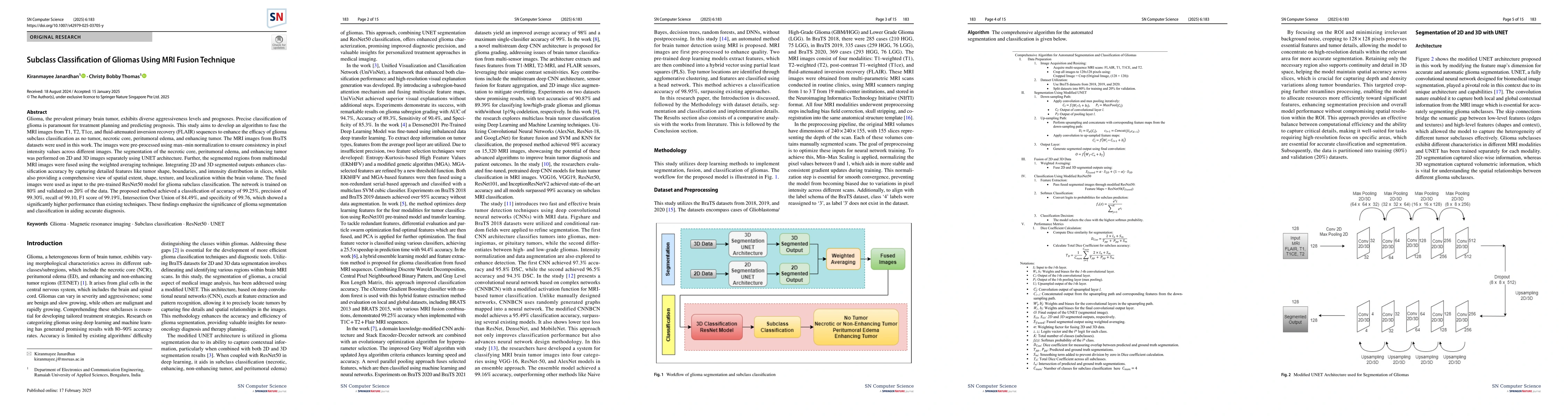
This study develops an algorithm to fuse MRI images (T1, T2, T1ce, FLAIR) using max-min normalization, UNET architecture for segmentation, and weighted averaging for feature fusion. The fused images are then classified using a pre-trained ResNet50 model.
Key Results — What are the main findings?
- The proposed method achieved a classification accuracy of 99.25%.
- Precision, recall, F1 score, and specificity were all high (99.30%, 99.10%, 99.19%, and 99.76% respectively).
- The performance significantly outperformed existing techniques.
Significance — Why does this research matter?
This research emphasizes the importance of accurate glioma segmentation and classification for effective treatment planning and prognosis prediction.
Technical Contribution — What is the technical contribution?
The innovative fusion of segmented 2D and 3D MRI data with ResNet50's pre-trained ImageNet model for enhanced glioma subclass classification.
Novelty — What is new about this work?
This approach leverages the complementary strengths of 2D and 3D MRI data, providing a comprehensive understanding and analysis of gliomas, surpassing previous methods in accuracy.
Limitations — What are the limitations of this study?
- The study did not address the influence of dataset variety on model effectiveness.
- Fixed weighting in data fusion may limit model adaptability to unique dataset characteristics.
Future Work — What did the authors propose for future work?
- Explore dynamic or adaptive weighting for better handling of diverse datasets.
- Investigate the model's adaptability to various clinical environments and populations.
Paper Details
How to Cite This Paper
@article{thomas2025subclass,
  title         = {Subclass Classification of Gliomas Using MRI Fusion Technique},
  author        = {Thomas, Christy Bobby and Janardhan, Kiranmayee},
  year          = {2025},
  eprint        = {2502.18775},
  archivePrefix = {arXiv},
  primaryClass  = {eess.IV},
  doi           = {10.1007/s42979-025-03705-y},
}Thomas, C., & Janardhan, K. (2025). Subclass Classification of Gliomas Using MRI Fusion Technique. arXiv. https://doi.org/10.1007/s42979-025-03705-yThomas, Christy Bobby, and Kiranmayee Janardhan. "Subclass Classification of Gliomas Using MRI Fusion Technique." arXiv, 2025, doi.org/10.1007/s42979-025-03705-y.PDF Preview
Citation Network
Current paper (gray), citations (green), references (blue)
Display is limited for performance on very large graphs.
Similar Papers
Found 4 papersCoronavirus (COVID-19) Classification using Deep Features Fusion and Ranking Technique
Mucahid Barstugan, Saban Ozturk, Umut Ozkaya
Glioma Classification using Multi-sequence MRI and Novel Wavelets-based Feature Fusion
Kiranmayee Janardhan, Christy Bobby Thomas
Image Feature Fusion of Hyperspectral Imaging and MRI for Automated Subtype Classification and Grading of Adult Diffuse Gliomas According to the 2021 WHO Criteria.
Lu, Jie, Li, Xiaoran, Cheng, Ye et al.
No citations found for this paper.
Comments (0)