Super-resolution diamond magnetic microscopy of superparamagnetic nanoparticles
Publication
Metrics
AI Quick Summary
This paper presents a super-resolution diamond magnetic microscopy technique using optically controlled NV centers to image magnetic fields from superparamagnetic nanoparticles, achieving a lateral resolution of ~100 nm and resolving individual magnetic dipoles in clusters. The method's enhanced magnetic feature amplitudes and lower background fluorescence open new avenues for nanoscale magnetic imaging.
Paper Preview
Abstract
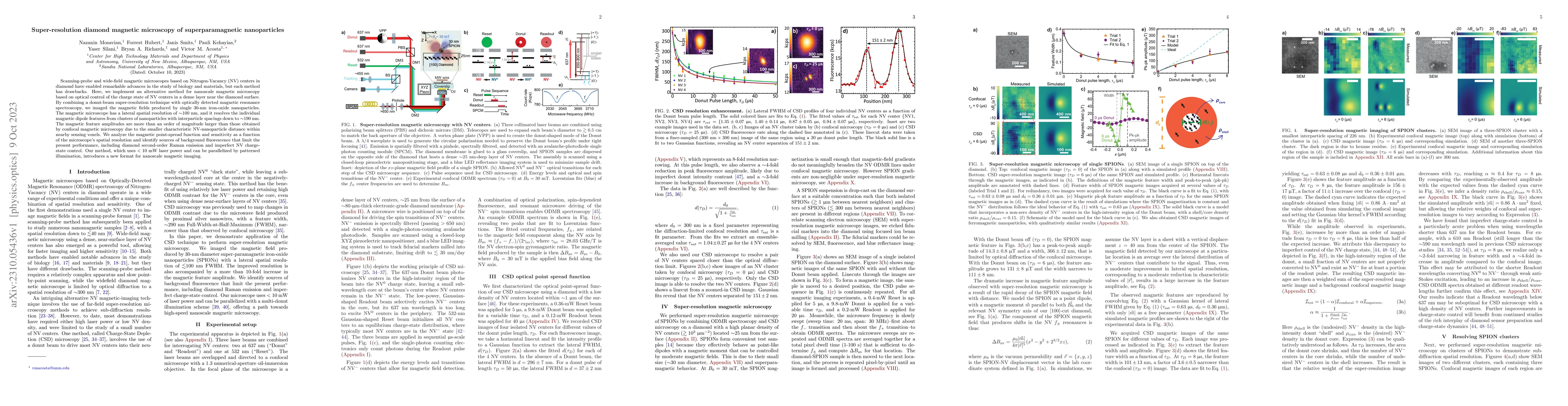
Scanning-probe and wide-field magnetic microscopes based on Nitrogen-Vacancy (NV) centers in diamond have enabled remarkable advances in the study of biology and materials, but each method has drawbacks. Here, we implement an alternative method for nanoscale magnetic microscopy based on optical control of the charge state of NV centers in a dense layer near the diamond surface. By combining a donut-beam super-resolution technique with optically detected magnetic resonance spectroscopy, we imaged the magnetic fields produced by single 30-nm iron-oxide nanoparticles. The magnetic microscope has a lateral spatial resolution of ~100 nm, and it resolves the individual magnetic dipole features from clusters of nanoparticles with interparticle spacings down to ~190 nm. The magnetic feature amplitudes are more than an order of magnitude larger than those obtained by confocal magnetic microscopy due to the smaller characteristic NV-nanoparticle distance within nearby sensing voxels. We analyze the magnetic point-spread function and sensitivity as a function of the microscope's spatial resolution and identify sources of background fluorescence that limit the present performance, including diamond second-order Raman emission and imperfect NV charge-state control. Our method, which uses less than 10 mW laser power and can be parallelized by patterned illumination, introduces a new format for nanoscale magnetic imaging.
AI Key Findings
Get AI-generated insights about this paper's methodology, results, significance, and more — seven facets brought into focus.
Impact
Paper Details
Authors
PDF Preview
Key Terms
Citation Network
Current paper (gray), citations (green), references (blue)
Display is limited for performance on very large graphs.
Discussion 0