ArxivLens
ArXiv physics.med-ph physics.bio-ph physics.optics

Super-resolution in vivo retinal imaging using structured illumination ophthalmoscopy

2007.16028 Published Oct 19, 2020
View PDF

Publication

Published: Oct 19, 2020
Categories: physics.med-ph, physics.bio-ph, physics.optics

Metrics

Source: ArXiv

AI Quick Summary

This paper presents advancements in structured illumination microscopy (SIM) for super-resolution in vivo retinal imaging, overcoming challenges such as motion, scattering, and optical aberrations. The use of adaptive optics and a specialized reconstruction algorithm enabled high-contrast super-resolution imaging of the human retina.

Quick Answers

What methodology did the authors use?

The research utilizes adaptive optics-corrected wide-field ophthalmoscopy with structured illumination (SIO) to achieve super-resolution and optical sectioning in the living retina, employing the BOSSA-SIM reconstruction algorithm. More in Methodology →

What are the key results?

SIO successfully demonstrated high contrast super-resolution in vivo imaging of the human retina, overcoming challenges such as sample motion, scattering, sample-induced optical aberrations, and low signal-to-noise ratio. — SIO images exhibited improved contrast and resolution compared to conventional wide-field ophthalmoscopy, particularly in visualizing foveal cones, which are crucial biomarkers for early detection of retinal degenerative diseases. More in Key Results →

Why is this work significant?

This research is significant as it introduces a novel, adaptive optics-corrected wide-field ophthalmoscope capable of super-resolution and optical sectioning in living retinas without distortion over large fields of view. This breakthrough could enhance retinal imaging, enabling better monitoring of retinal diseases and improving diagnostic accuracy. More in Significance →

What are the main limitations?

The resolution improvement is currently limited by the poor signal-to-noise ratio of raw images, which do not reach the diffraction limit due to noise. — The method relies on specific hardware and algorithms, which may not be readily accessible or applicable to all imaging setups. More in Limitations →

Paper Preview

Abstract

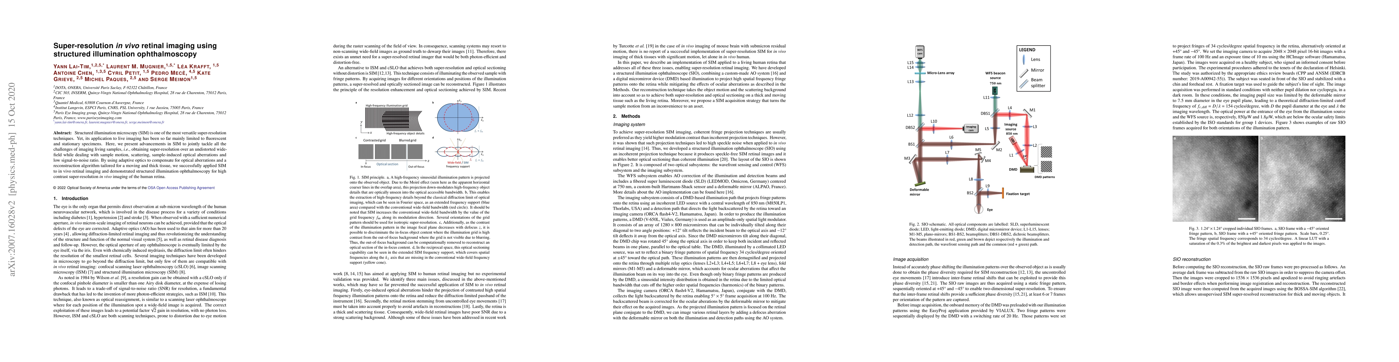
Structured illumination microscopy (SIM) is one of the most versatile super-resolution techniques. Yet, its application to live imaging has been so far mainly limited to fluorescent and stationary specimens. Here, we present advancements in SIM to jointly tackle all the challenges of imaging living samples, i.e., obtaining super-resolution over an undistorted wide-field while dealing with sample motion, scattering, sample-induced optical aberrations and low signal-to-noise ratio. By using adaptive optics to compensate for optical aberrations and a reconstruction algorithm tailored for a moving and thick tissue, we successfully applied SIM to in vivo retinal imaging and demonstrated structured illumination ophthalmoscopy for high contrast super-resolution in vivo imaging of the human retina.

Key Findings, in focus

Seven facets of this paper, analysed and brought into focus by AI.

AI-generated · verify against source Generated Sep 02, 2025
In focus · one-line synthesis

This research is significant as it introduces a novel, adaptive optics-corrected wide-field ophthalmoscope capable of super-resolution and optical sectioning in living retinas without distortion over large fields of view.

01
MethodologyHow they did it

The research utilizes adaptive optics-corrected wide-field ophthalmoscopy with structured illumination (SIO) to achieve super-resolution and optical sectioning in the living retina, employing the BOSSA-SIM reconstruction algorithm.

02
ResultsWhat they found
  • SIO successfully demonstrated high contrast super-resolution in vivo imaging of the human retina, overcoming challenges such as sample motion, scattering, sample-induced optical aberrations, and low signal-to-noise ratio.
  • SIO images exhibited improved contrast and resolution compared to conventional wide-field ophthalmoscopy, particularly in visualizing foveal cones, which are crucial biomarkers for early detection of retinal degenerative diseases.
  • The SIO method allowed for more accurate cone density measurements over a wider range of retinal eccentricities, with distortion-free images suitable for longitudinal studies.
03
SignificanceWhy it matters

This research is significant as it introduces a novel, adaptive optics-corrected wide-field ophthalmoscope capable of super-resolution and optical sectioning in living retinas without distortion over large fields of view. This breakthrough could enhance retinal imaging, enabling better monitoring of retinal diseases and improving diagnostic accuracy.

04
Technical ContributionsWhat's new technically

The BOSSA-SIM reconstruction algorithm, which features five main aspects: an imaging model accounting for retinal shifts, a 2-layer object model distinguishing in-focus signal from out-of-focus contributions, robustness to noise through a Bayesian framework, automatic adjustment of reconstruction hyper-parameters, and a positivity constraint on the reconstructed object.

05
NoveltyWhat sets it apart

This work stands out by combining super-resolution and distortion-free acquisition in wide-field retinal imaging, which has been a challenge in the past. It provides a generic strategy for achieving super-resolution and optical sectioning in various biomedical imaging applications despite sample motion, scattering, and aberrations.

06
LimitationsWhat to be cautious about
  • The resolution improvement is currently limited by the poor signal-to-noise ratio of raw images, which do not reach the diffraction limit due to noise.
  • The method relies on specific hardware and algorithms, which may not be readily accessible or applicable to all imaging setups.
07
Future WorkWhere it goes next
  • Ongoing developments aim to improve the signal-to-noise ratio of raw images to further increase resolution enhancement enabled by SIO.
  • Exploring the application of this technique in various biomedical imaging scenarios with sample motion, scattering, and aberrations.

Impact

36
References

Paper Details

Paper ID: 2007.16028
License: http://arxiv.org/licenses/nonexclusive-distrib/1.0/
Comments: 14 pages, 8 figures, manuscript under consideration for a peer-reviewed journal
Categories:
physics.med-ph physics.bio-ph physics.optics

How to Cite This Paper

Reference manager: RIS EndNote

Key Terms

super resolution (0.315)
imaging (0.262)
illumination (0.259)
super (0.251)
sim (0.228)
structured (0.209)
resolution (0.200)
optical (0.157)

Citation Network

Current paper (gray), citations (green), references (blue)

Display is limited for performance on very large graphs.

Current Paper
Citations
References
Click to view

Related Papers

4 found


Discussion 0

This website uses cookies to ensure you get the best experience. Learn more