Publication
Metrics
AI Quick Summary
This paper presents a genetically optimized apochromatic super-oscillatory lens (SOL) that enables multicolor super-resolution fluorescence microscopy with extended depth-of-focus, achieving sub-diffraction-limit imaging across multiple wavelengths and minimizing chromatic aberrations. The designed lens facilitates three-dimensional imaging of neuronal structures without complex sample positioning.
Paper Preview
Abstract
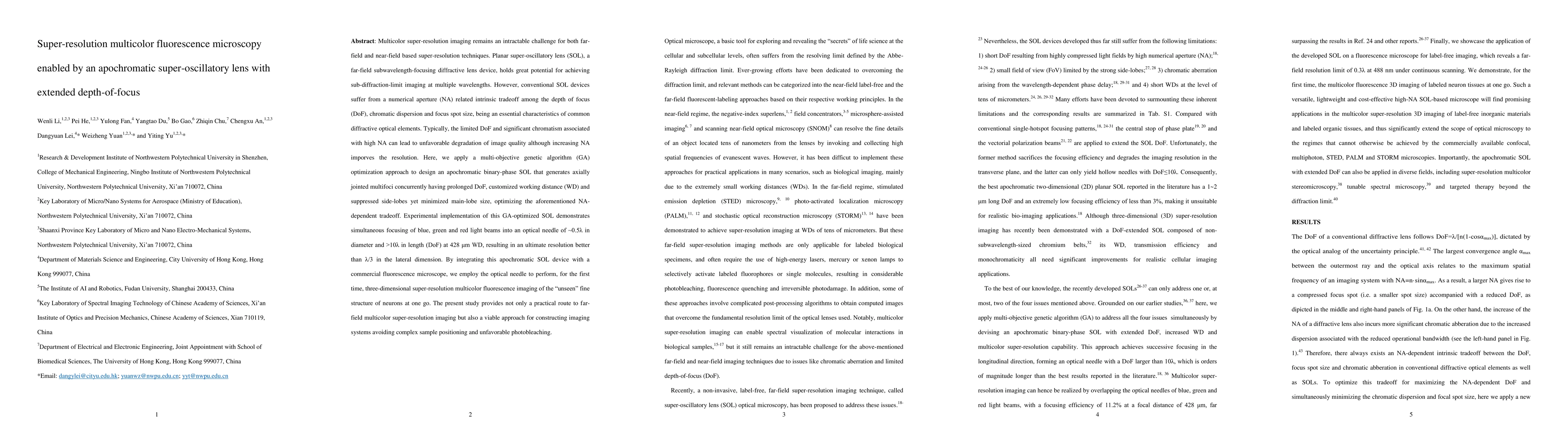
Multicolor super-resolution imaging remains an intractable challenge for both far-field and near-field based super-resolution techniques. Planar super-oscillatory lens (SOL), a far-field subwavelength-focusing diffractive lens device, holds great potential for achieving sub-diffraction-limit imaging at multiple wavelengths. However, conventional SOL devices suffer from a numerical aperture (NA) related intrinsic tradeoff among the depth of focus (DoF), chromatic dispersion and focus spot size, being an essential characteristics of common diffractive optical elements. Typically, the limited DoF and significant chromatism associated with high NA can lead to unfavorable degradation of image quality although increasing NA imporves the resolution. Here, we apply a multi-objective genetic algorithm (GA) optimization approach to design an apochromatic binary-phase SOL that generates axially jointed multifoci concurrently having prolonged DoF, customized working distance (WD) and suppressed side-lobes yet minimized main-lobe size, optimizing the aforementioned NA-dependent tradeoff. Experimental implementation of this GA-optimized SOL demonstrates simultaneous focusing of blue, green and red light beams into an optical needle half of the incident wavelength in diameter at 428 um WD, resulting in an ultimate resolution better than one third of the incident wavelength in the lateral dimension. By integrating this apochromatic SOL device with a commercial fluorescence microscope, we employ the optical needle to perform, for the first time, three-dimensional super-resolution multicolor fluorescence imaging of the unseen fine structure of neurons at one go. The present study provides not only a practical route to far-field multicolor super-resolution imaging but also a viable approach for constructing imaging systems avoiding complex sample positioning and unfavorable photobleaching.
AI Key Findings
Get AI-generated insights about this paper's methodology, results, significance, and more — seven facets brought into focus.
Impact
Paper Details
Authors
PDF Preview
Key Terms
Citation Network
Current paper (gray), citations (green), references (blue)
Display is limited for performance on very large graphs.
Discussion 0