Publication
Metrics
Quick Actions
AI Quick Summary
This paper introduces Deep Ultrasound Localization Microscopy (Deep-ULM), a deep learning-based method to achieve super-resolution vascular imaging in high-density contrast-enhanced ultrasound data. Deep-ULM uses a convolutional neural network to accurately localize microbubbles, significantly improving resolution and reducing acquisition times.
Quick Answers
What is "Super-resolution Ultrasound Localization Microscopy through Deep Learning" about?
This paper introduces Deep Ultrasound Localization Microscopy (Deep-ULM), a deep learning-based method to achieve super-resolution vascular imaging in high-density contrast-enhanced ultrasound data. Deep-ULM uses a convolutional neural network to accurately localize microbubbles, significantly improving resolution and reducing acquisition times.
What methodology did the authors use?
The research introduces Deep Ultrasound Localization Microscopy (Deep-ULM), a deep learning-based method that leverages convolutional neural networks for precise super-resolution vascular imaging in high-density ultrasound contrast agent scenarios. It synthesizes training data online and trains an end-to-end fully convolutional neural network for robust inference under various imaging conditions. More in Methodology →
What are the key results?
Deep-ULM achieves super-resolution imaging with high contrast-agent densities, both in-silico and in-vivo. — The method resolves approximately 70 high-resolution patches (128x128 pixels) per second on a standard PC, with GPU computation increasing this to 1250 patches per second, making it suitable for real-time applications. More in Key Results →
Why is this work significant?
This research is significant as it addresses the limitations of traditional ultrasound localization microscopy by enabling precise super-resolution imaging in high-density contrast agent scenarios, which is crucial for reducing acquisition times and improving the utility of ultrasound imaging in clinical settings. More in Significance →
What are the main limitations?
The current implementation is limited to 2D imaging, with future work aiming to extend it to 3D for broader applicability. — The method was not trained with structural priors on vascular architecture or microbubble dynamics, which could potentially enhance image fidelity in future iterations. More in Limitations →
Abstract
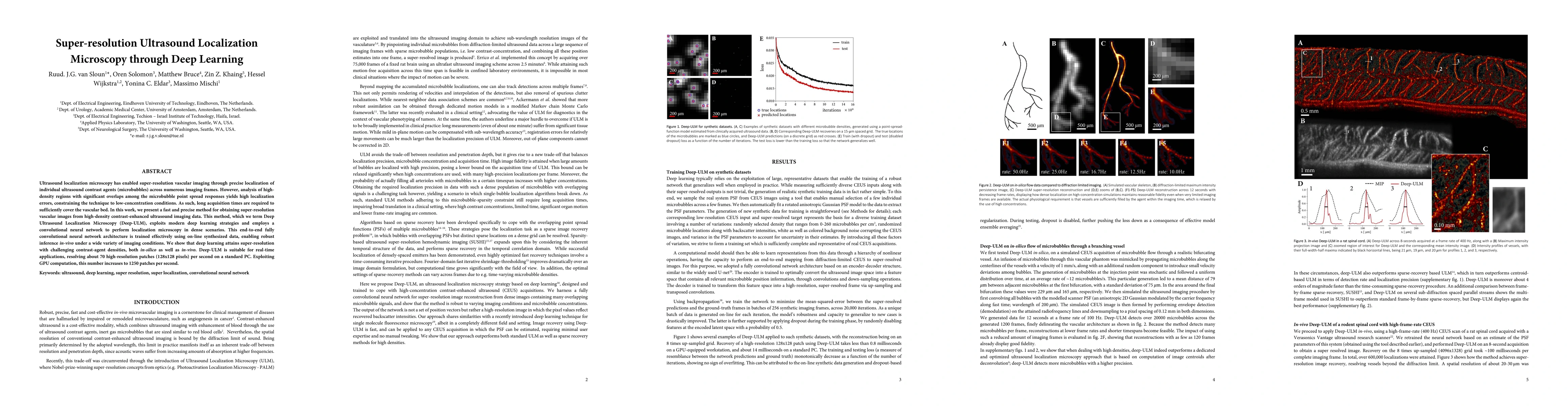
Ultrasound localization microscopy has enabled super-resolution vascular imaging through precise localization of individual ultrasound contrast agents (microbubbles) across numerous imaging frames. However, analysis of high-density regions with significant overlaps among the microbubble point spread responses yields high localization errors, constraining the technique to low-concentration conditions. As such, long acquisition times are required to sufficiently cover the vascular bed. In this work, we present a fast and precise method for obtaining super-resolution vascular images from high-density contrast-enhanced ultrasound imaging data. This method, which we term Deep Ultrasound Localization Microscopy (Deep-ULM), exploits modern deep learning strategies and employs a convolutional neural network to perform localization microscopy in dense scenarios. This end-to-end fully convolutional neural network architecture is trained effectively using on-line synthesized data, enabling robust inference in-vivo under a wide variety of imaging conditions. We show that deep learning attains super-resolution with challenging contrast-agent densities, both in-silico as well as in-vivo. Deep-ULM is suitable for real-time applications, resolving about 70 high-resolution patches (128x128 pixels) per second on a standard PC. Exploiting GPU computation, this number increases to 1250 patches per second.
AI Key Findings
Generated Sep 02, 2025
Methodology — What approach did the authors take?
The research introduces Deep Ultrasound Localization Microscopy (Deep-ULM), a deep learning-based method that leverages convolutional neural networks for precise super-resolution vascular imaging in high-density ultrasound contrast agent scenarios. It synthesizes training data online and trains an end-to-end fully convolutional neural network for robust inference under various imaging conditions.
Key Results — What are the main findings?
- Deep-ULM achieves super-resolution imaging with high contrast-agent densities, both in-silico and in-vivo.
- The method resolves approximately 70 high-resolution patches (128x128 pixels) per second on a standard PC, with GPU computation increasing this to 1250 patches per second, making it suitable for real-time applications.
- Deep-ULM outperforms traditional ULM and other ULM variants (centroid localization, sparse recovery) in terms of detection rate and localization precision, especially at high microbubble densities.
- Initial in-vivo testing on a human prostate demonstrated the feasibility of Deep-ULM with a standard ultrasound system, achieving real-time inference rates above 20 Hz.
Significance — Why does this research matter?
This research is significant as it addresses the limitations of traditional ultrasound localization microscopy by enabling precise super-resolution imaging in high-density contrast agent scenarios, which is crucial for reducing acquisition times and improving the utility of ultrasound imaging in clinical settings.
Technical Contribution — What is the technical contribution?
The main technical contribution is the development of Deep-ULM, a deep learning-based method that uses a convolutional neural network to perform localization microscopy in dense ultrasound contrast agent scenarios, significantly improving precision and enabling real-time processing.
Novelty — What is new about this work?
The novelty of this work lies in the application of deep learning techniques to ultrasound localization microscopy, enabling precise super-resolution imaging in high-density contrast agent conditions, which was previously constrained by high localization errors and long acquisition times.
Limitations — What are the limitations of this study?
- The current implementation is limited to 2D imaging, with future work aiming to extend it to 3D for broader applicability.
- The method was not trained with structural priors on vascular architecture or microbubble dynamics, which could potentially enhance image fidelity in future iterations.
Future Work — What did the authors propose for future work?
- Investigate incorporating structural priors to improve image fidelity and generalization to pathological conditions.
- Explore the extension of Deep-ULM to 3D imaging for more comprehensive applications.
Paper Details
How to Cite This Paper
@article{anon2018super,
title = {Super-resolution Ultrasound Localization Microscopy through Deep
Learning},
year = {2018},
eprint = {1804.07661},
archivePrefix = {arXiv},
primaryClass = {eess.SP},
}
(2018). Super-resolution Ultrasound Localization Microscopy through Deep
Learning. arXiv. https://arxiv.org/abs/1804.07661
"Super-resolution Ultrasound Localization Microscopy through Deep
Learning." arXiv, 2018, arxiv.org/abs/1804.07661.
PDF Preview
Key Terms
Citation Network
Current paper (gray), citations (green), references (blue)
Display is limited for performance on very large graphs.
Similar Papers
Found 4 papersLearning Super-Resolution Ultrasound Localization Microscopy from Radio-Frequency Data
Christopher Hahne, Georges Chabouh, Olivier Couture et al.
Advancing biological super-resolution microscopy through deep learning: a brief review
Tianjie Yang, Yaoru Luo, Ge Yang et al.
Ensemble Learning for Microbubble Localization in Super-Resolution Ultrasound
Sepideh K. Gharamaleki, Brandon Helfield, Hassan Rivaz
A Deep Learning Framework for Spatiotemporal Ultrasound Localization Microscopy
Léo Milecki, Jonathan Porée, Hatim Belgharbi et al.
| Title | Authors | Year | Actions |
|---|
Comments (0)