Summary
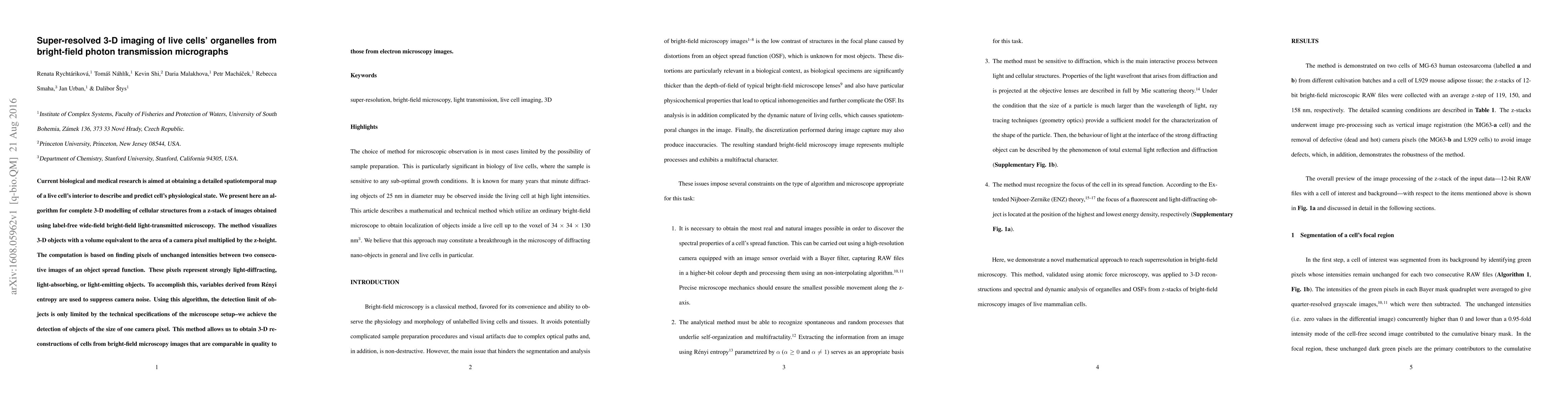
Current biological and medical research is aimed at obtaining a detailed spatiotemporal map of a live cell's interior to describe and predict cell's physiological state. We present here an algorithm for complete 3-D modelling of cellular structures from a z-stack of images obtained using label-free wide-field bright-field light-transmitted microscopy. The method visualizes 3-D objects with a volume equivalent to the area of a camera pixel multiplied by the z-height. The computation is based on finding pixels of unchanged intensities between two consecutive images of an object spread function. These pixels represent strongly light-diffracting, light-absorbing, or light-emitting objects. To accomplish this, variables derived from R\'{e}nyi entropy are used to suppress camera noise. Using this algorithm, the detection limit of objects is only limited by the technical specifications of the microscope setup--we achieve the detection of objects of the size of one camera pixel. This method allows us to obtain 3-D reconstructions of cells from bright-field microscopy images that are comparable in quality to those from electron microscopy images.
AI Key Findings
Get AI-generated insights about this paper's methodology, results, and significance.
Paper Details
PDF Preview
Key Terms
Citation Network
Current paper (gray), citations (green), references (blue)
Display is limited for performance on very large graphs.
Similar Papers
Found 4 papersIn silico prediction of cellular organelles from computationally super-resolved (SR) phase-modulated optical micrographs
Sharma, A., Kaderuppan, S. S., Saifuddin, M. R. et al.
Super-resolution Live-cell Fluorescence Lifetime Imaging
Wolfgang Hübner, Thomas Juffmann, Thomas Huser et al.
| Title | Authors | Year | Actions |
|---|
Comments (0)