Publication
Metrics
AI Quick Summary
This paper presents a fully automatic framework for reconstructing high-resolution, large-field-of-view retinal images from multiple low-resolution frames using a low-cost, mobile camera. The method combines super-resolved image fusion and mosaicing to achieve clinically useful results.
Paper Preview
Abstract
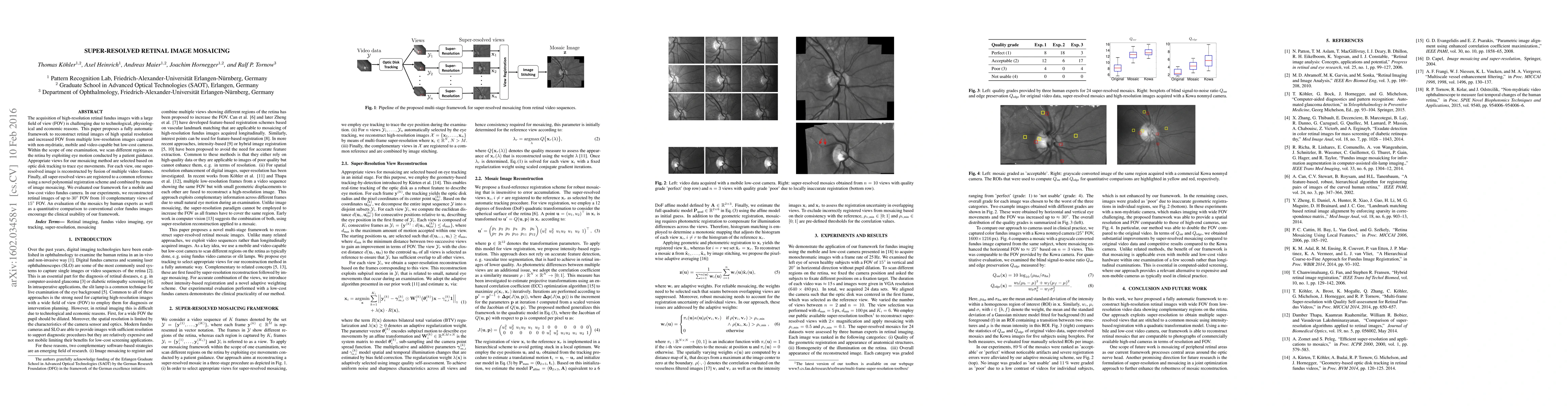
The acquisition of high-resolution retinal fundus images with a large field of view (FOV) is challenging due to technological, physiological and economic reasons. This paper proposes a fully automatic framework to reconstruct retinal images of high spatial resolution and increased FOV from multiple low-resolution images captured with non-mydriatic, mobile and video-capable but low-cost cameras. Within the scope of one examination, we scan different regions on the retina by exploiting eye motion conducted by a patient guidance. Appropriate views for our mosaicing method are selected based on optic disk tracking to trace eye movements. For each view, one super-resolved image is reconstructed by fusion of multiple video frames. Finally, all super-resolved views are registered to a common reference using a novel polynomial registration scheme and combined by means of image mosaicing. We evaluated our framework for a mobile and low-cost video fundus camera. In our experiments, we reconstructed retinal images of up to 30{\deg} FOV from 10 complementary views of 15{\deg} FOV. An evaluation of the mosaics by human experts as well as a quantitative comparison to conventional color fundus images encourage the clinical usability of our framework.
AI Key Findings
Get AI-generated insights about this paper's methodology, results, significance, and more — seven facets brought into focus.
Impact
Paper Details
PDF Preview
Key Terms
Citation Network
Current paper (gray), citations (green), references (blue)
Display is limited for performance on very large graphs.
Discussion 0