Publication
Metrics
AI Quick Summary
This paper proposes a method using superpixel segmentation (SLIC and QS) to identify five GI diseases in Wireless Capsule Endoscopy images, followed by SVM classification based on texture and color features. The method achieves high accuracy (~92%) and is significantly faster with SLIC compared to QS.
Paper Preview
Abstract
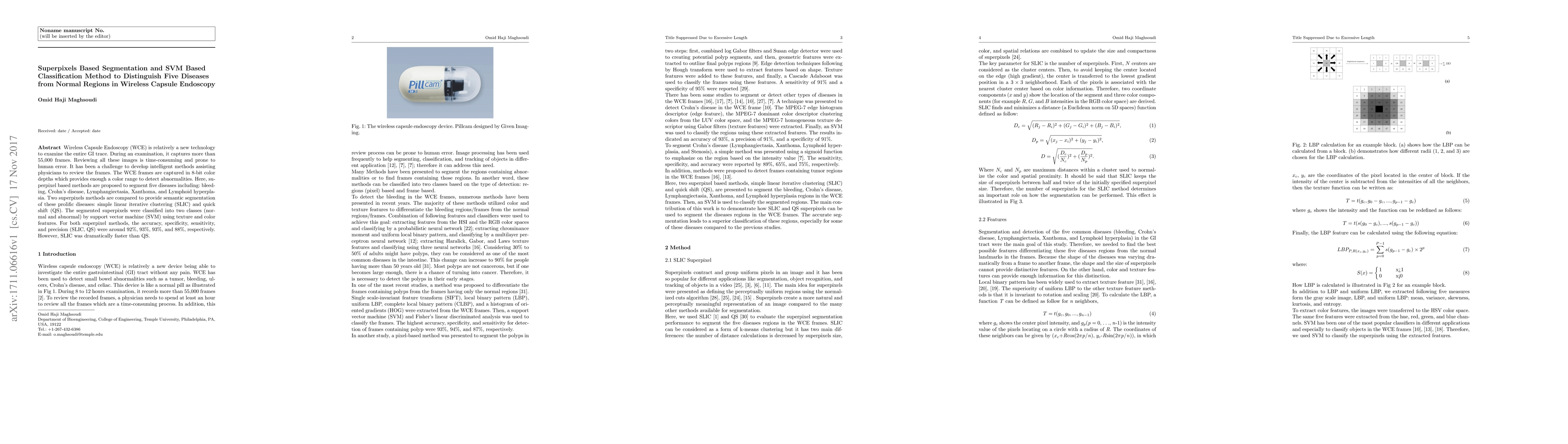
Wireless Capsule Endoscopy (WCE) is relatively a new technology to examine the entire GI trace. During an examination, it captures more than 55,000 frames. Reviewing all these images is time-consuming and prone to human error. It has been a challenge to develop intelligent methods assisting physicians to review the frames. The WCE frames are captured in 8-bit color depths which provides enough a color range to detect abnormalities. Here, superpixel based methods are proposed to segment five diseases including: bleeding, Crohn's disease, Lymphangiectasia, Xanthoma, and Lymphoid hyperplasia. Two superpixels methods are compared to provide semantic segmentation of these prolific diseases: simple linear iterative clustering (SLIC) and quick shift (QS). The segmented superpixels were classified into two classes (normal and abnormal) by support vector machine (SVM) using texture and color features. For both superpixel methods, the accuracy, specificity, sensitivity, and precision (SLIC, QS) were around 92%, 93%, 93%, and 88%, respectively. However, SLIC was dramatically faster than QS.
AI Key Findings
Get AI-generated insights about this paper's methodology, results, significance, and more — seven facets brought into focus.
Impact
Paper Details
PDF Preview
Key Terms
Citation Network
Current paper (gray), citations (green), references (blue)
Display is limited for performance on very large graphs.
Discussion 0