Surface-guided computing to analyze subcellular morphology and membrane-associated signals in 3D
Publication
Metrics
AI Quick Summary
This paper introduces u-Unwrap3D, a framework for analyzing subcellular morphology and membrane-associated signals in 3D by remapping complex cell surfaces into lower dimensions, enabling quantitative spatiotemporal analyses of cellular processes such as polymer recruitment and ruffle movement.
Paper Preview
Abstract
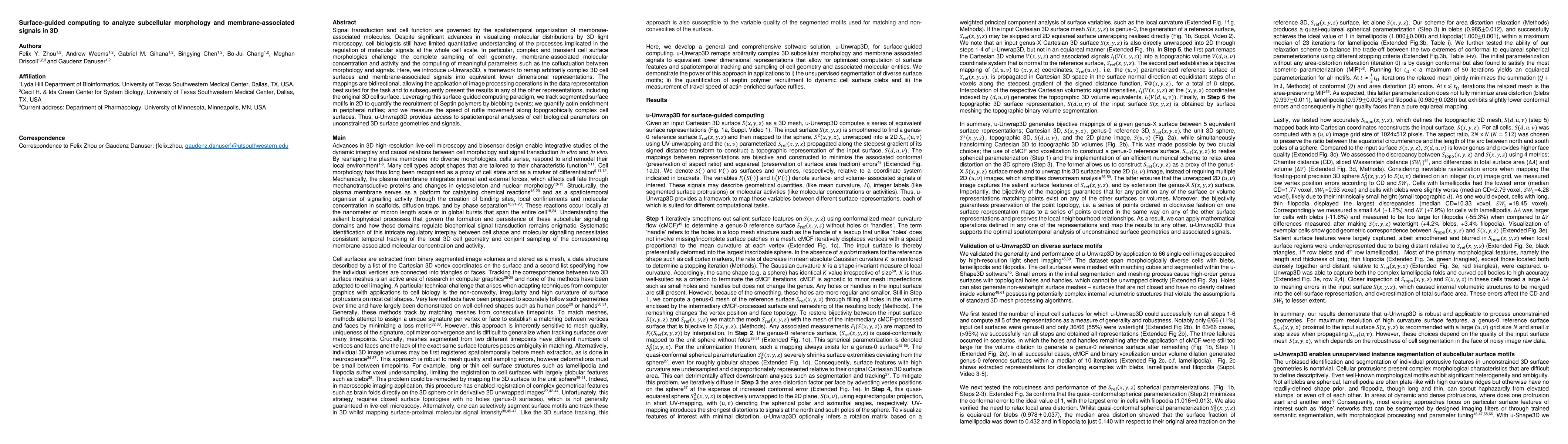
Signal transduction and cell function are governed by the spatiotemporal organization of membrane-associated molecules. Despite significant advances in visualizing molecular distributions by 3D light microscopy, cell biologists still have limited quantitative understanding of the processes implicated in the regulation of molecular signals at the whole cell scale. In particular, complex and transient cell surface morphologies challenge the complete sampling of cell geometry, membrane-associated molecular concentration and activity and the computing of meaningful parameters such as the cofluctuation between morphology and signals. Here, we introduce u-Unwrap3D, a framework to remap arbitrarily complex 3D cell surfaces and membrane-associated signals into equivalent lower dimensional representations. The mappings are bidirectional, allowing the application of image processing operations in the data representation best suited for the task and to subsequently present the results in any of the other representations, including the original 3D cell surface. Leveraging this surface-guided computing paradigm, we track segmented surface motifs in 2D to quantify the recruitment of Septin polymers by blebbing events; we quantify actin enrichment in peripheral ruffles; and we measure the speed of ruffle movement along topographically complex cell surfaces. Thus, u-Unwrap3D provides access to spatiotemporal analyses of cell biological parameters on unconstrained 3D surface geometries and signals.
AI Key Findings
Get AI-generated insights about this paper's methodology, results, significance, and more — seven facets brought into focus.
Impact
Paper Details
Authors
PDF Preview
Key Terms
Citation Network
Current paper (gray), citations (green), references (blue)
Display is limited for performance on very large graphs.
Discussion 0