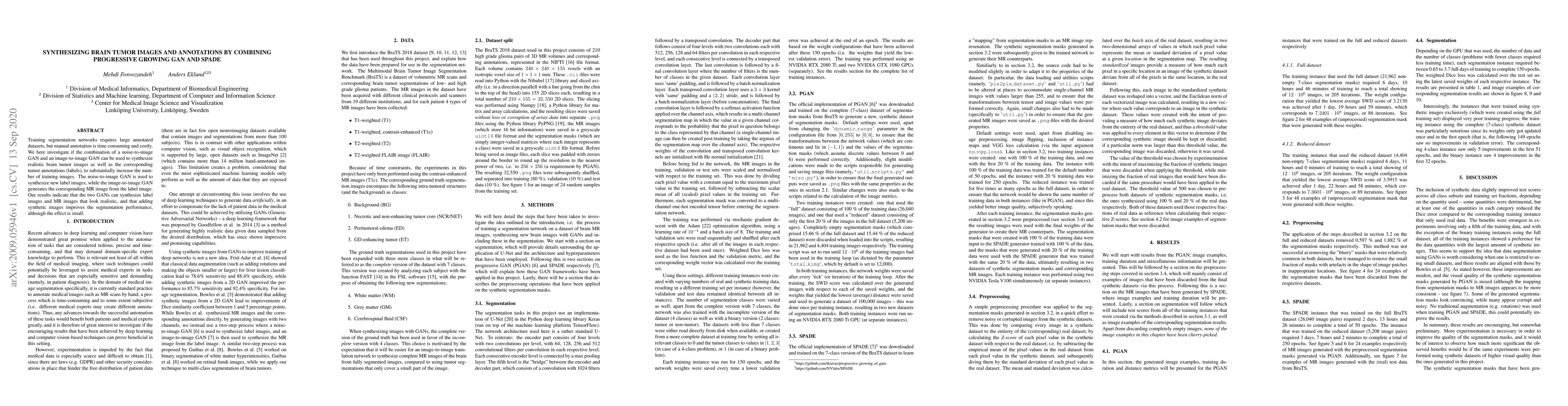
ArxivLens

Quick Summary:

This paper explores the use of a combination of progressive growing GAN and SPADE to synthesize realistic brain tumor images and corresponding annotations, aiming to augment training datasets for segmentation networks. The results show that the synthesized images and annotations are realistic and lead to a small but positive improvement in segmentation performance.

arXiv Id: 2009.05946
Date Published: 2020-09-15
Date Updated: 2020-09-15

Categories:

Key Terms (TF-IDF):

gan (0.367)
gan (0.353)
image (0.302)
synthesize (0.284)
images (0.284)
image (0.284)
label (0.282)
mr (0.275)
tumor (0.267)
images (0.263)
label (0.258)
segmentation (0.225)
segmentation (0.204)
realistic (0.197)
realistic (0.186)
noise (0.169)
noise (0.160)
training (0.159)
annotations (0.154)
annotated (0.153)
License: http://creativecommons.org/licenses/by/4.0/

Summary:

Training segmentation networks requires large annotated datasets, but manual annotation is time consuming and costly. We here investigate if the combination of a noise-to-image GAN and an image-to-image GAN can be used to synthesize realistic brain tumor images as well as the corresponding tumor annotations (labels), to substantially increase the number of training images. The noise-to-image GAN is used to synthesize new label images, while the image-to-image GAN generates the corresponding MR image from the label image. Our results indicate that the two GANs can synthesize label images and MR images that look realistic, and that adding synthetic images improves the segmentation performance, although the effect is small.

Citation & Reference Network Visualization

This graph shows how this paper is connected to other research. The current paper is shown in dark gray, papers that cite it are in green, and papers it references are in blue. You can:

Current Paper
Papers citing this work (Citations)
Papers cited by this work (References)

• Hover over any node to see the paper title

• Click a node to view that paper's details

• Drag nodes to rearrange the graph

• Use the zoom controls (bottom right) to zoom in/out

Citations and References

Title Authors Year Actions
Investigating the Use of Gan in the Classification of Brain Tumors
Michaël Jordan Ah-Shee-Tee, Rousitha Chikhuri, Zahra Mungloo-Dilmohamud
2023
View Paper
Evaluating synthetic neuroimaging data augmentation for automatic brain tumour segmentation with a deep fully-convolutional network
Fawad Asadi, Thanate Angsuwatanakul, Jamie A O’Reilly
2023
View Paper
A 3D generative model of pathological multi-modal MR images and segmentations
Virginia Fernandez, W. H. Pinaya, Pedro Borges, M. Graham, Tom Vercauteren, M. J. Cardoso
2023
View Paper
MIPR: Automatic Segmentation Annotation of Medical Images via Generative model
Haiming Zhu, Pingping Dai, Hao Wang, Xiang Qian
2023
View Paper
Brain tumor segmentation using synthetic MR images - A comparison of GANs and diffusion models
M. U. Akbar, Måns Larsson, Anders Eklund
2023
View Paper
Does an ensemble of GANs lead to better performance when training segmentation networks with synthetic images?
Maans Larsson, M. U. Akbar, A. Eklund
2022
View Paper
Style Transfer with Bio-realistic Appearance Manipulation for Skin-tone Inclusive rPPG
Yunhao Ba, Zhen Wang, Kerim Doruk Karinca, Oyku Deniz Bozkurt, A. Kadambi
2022
View Paper
Generation of Synthetic Rat Brain MRI Scans with a 3D Enhanced Alpha Generative Adversarial Network
André Ferreira, R. Magalhães, S. Mériaux, Victor Alves
2022
View Paper
Toward Sharing Brain Images: Differentially Private TOF-MRA Images With Segmentation Labels Using Generative Adversarial Networks
Tabea Kossen, Manuel A. Hirzel, V. Madai, Franziska Boenisch, A. Hennemuth, Kristian Hildebrand, S. Pokutta, Kartikey Sharma, A. Hilbert, J. Sobesky, Ivana Galinovic, A. Khalil, J. Fiebach, D. Frey
2022
View Paper
MIPR: Automatic Annotation of Medical Images with Pixel Rearrangement
Pingping Dai, Haiming Zhu, Shuang Ge, Ruihan Zhang, Xiang Qian, Xi Li, Kehong Yuan
2022
View Paper
Soft-CP: A Credible and Effective Data Augmentation for Semantic Segmentation of Medical Lesions
Pingping Dai, Licong Dong, Ruihan Zhang, Haiming Zhu, Jie Wu, Kehong Yuan
2022
View Paper
Generating 3D TOF-MRA volumes and segmentation labels using generative adversarial networks
Pooja Subramaniam, Tabea Kossen, K. Ritter, A. Hennemuth, Kristian Hildebrand, A. Hilbert, J. Sobesky, Michelle Livne, Ivana Galinovic, A. Khalil, J. Fiebach, D. Frey, V. Madai
2022
View Paper
Generation of Synthetic Rat Brain MRI scans with a 3D Enhanced Alpha-GAN
André Ferreira, R. Magalhães, S. Mériaux, Victor Alves
2021
View Paper
Data synthesis and adversarial networks: A review and meta-analysis in cancer imaging
Richard Osuala, Kaisar Kushibar, Lidia Garrucho, Akis Linardos, Zuzanna Szafranowska, Stefan Klein, Ben Glocker, Oliver Díaz, K. Lekadir
2021
View Paper
Anonymization of labeled TOF-MRA images for brain vessel segmentation using generative adversarial networks
Tabea Kossen, Pooja Subramaniam, V. Madai, A. Hennemuth, Kristian Hildebrand, A. Hilbert, J. Sobesky, Michelle Livne, Ivana Galinovic, A. Khalil, J. Fiebach, D. Frey
2020
View Paper
A Review of Generative Adversarial Networks in Cancer Imaging: New Applications, New Solutions
Richard Osuala, Kaisar Kushibar, Lidia Garrucho, Akis Linardos, Zuzanna Szafranowska, S. Klein, Ben Glocker, Oliver Díaz, Karim Lekadir
2021
View Paper
BrainPixGAN: Generating Intraoperative MR with Mask-Based Generative Networks
Ayşe Gül Eker, Meltem Kurt, Pehlivanoglu, Ayşe Gül Eker, Meltem Kurt Pehlivano˘glu, Nevcihan Duru, Tolga Turan D¨undar
View Paper

Similar Papers

Automatic brain tumor segmentation in 2D intra-operative ultrasound images using MRI tumor annotations

Ingerid Reinertsen, Ole Solheim, Ragnhild Holden Helland, Mathilde Faanes

Published: 2024-11-21

RESECT-SEG: Open access annotations of intra-operative brain tumor ultrasound images

Yiming Xiao, Hassan Rivaz, Ingerid Reinertsen, Ole Solheim, Sandrine De Ribaupierre, Matthieu Chabanas, Bahareh Behboodi, Francois-Xavier Carton, Bodil K. R. Munkvold

Published: 2022-07-18

Comments (0)

This website uses cookies to ensure you get the best experience. Learn more