ArxivLens
ArXiv cs.CV

T-Gated Adapter: A Lightweight Temporal Adapter for Vision-Language Medical Segmentation

Pranjal Khadka

2604.08167 Published Apr 9, 2026
View PDF

Publication

Published: Apr 09, 2026
Categories: cs.CV

Metrics

Source: ArXiv

Quick Answers

What methodology did the authors use?

Fine‑tune a CLIPSeg 2‑D vision‑language model on 30 labeled CT volumes, augmenting it with a lightweight temporal‑spatial adapter that injects a 5‑slice context via a temporal transformer, spatial context block, and adaptive gate. More in Methodology →

What are the key results?

+0.206 Dice improvement over baseline on FLARE22 (mean Dice 0.704 vs 0.497) — +0.210 and +0.230 Dice gains on zero‑shot BTCV and AMOS22 cross‑domain CT benchmarks More in Key Results →

Why is this work significant?

Demonstrates that 2‑D vision‑language models can be extended to volumetric medical segmentation with minimal data, achieving robust cross‑domain and cross‑modality performance and potentially reducing the need for dense 3‑D annotations in clinical practice. More in Significance →

What are the main limitations?

Fixed 5‑slice context window ignores varying slice spacing across CT acquisitions — Downsampling CT slices to 352×352 discards fine spatial detail, limiting performance on small organs More in Limitations →

Paper Preview

Abstract

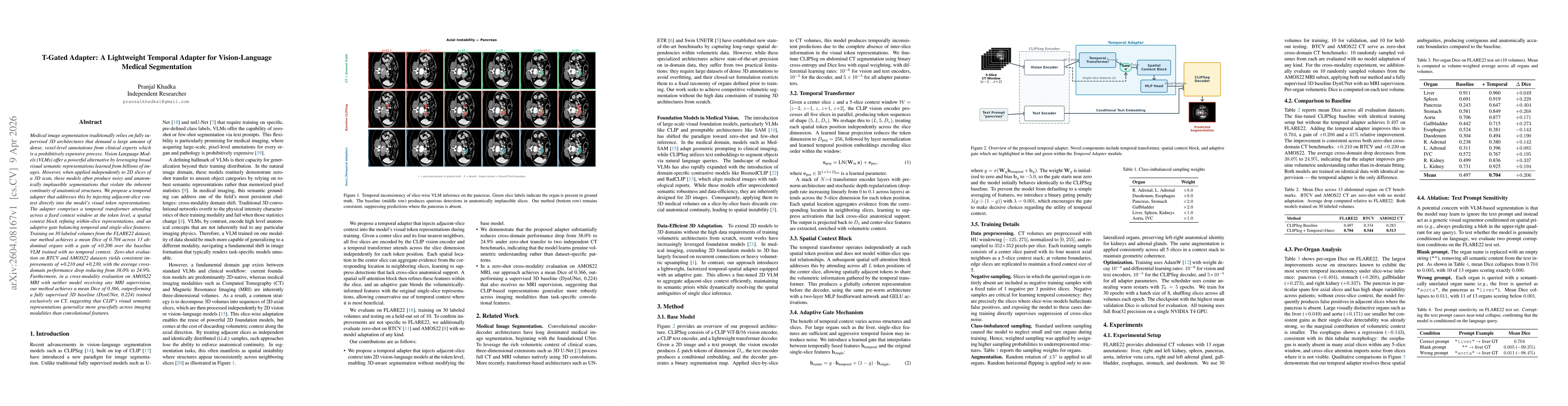
Medical image segmentation traditionally relies on fully supervised 3D architectures that demand a large amount of dense, voxel-level annotations from clinical experts which is a prohibitively expensive process. Vision Language Models (VLMs) offer a powerful alternative by leveraging broad visual semantic representations learned from billions of images. However, when applied independently to 2D slices of a 3D scan, these models often produce noisy and anatomically implausible segmentations that violate the inherent continuity of anatomical structures. We propose a temporal adapter that addresses this by injecting adjacent-slice context directly into the model's visual token representations. The adapter comprises a temporal transformer attending across a fixed context window at the token level, a spatial context block refining within-slice representations, and an adaptive gate balancing temporal and single-slice features. Training on 30 labeled volumes from the FLARE22 dataset, our method achieves a mean Dice of 0.704 across 13 abdominal organs with a gain of +0.206 over the baseline VLM trained with no temporal context. Zero-shot evaluation on BTCV and AMOS22 datasets yields consistent improvements of +0.210 and +0.230, with the average cross-domain performance drop reducing from 38.0% to 24.9%. Furthermore, in a cross-modality evaluation on AMOS22 MRI with neither model receiving any MRI supervision, our method achieves a mean Dice of 0.366, outperforming a fully supervised 3D baseline (DynUNet, 0.224) trained exclusively on CT, suggesting that CLIP's visual semantic representations generalize more gracefully across imaging modalities than convolutional features.

Key Findings, in focus

Seven facets of this paper, analysed and brought into focus by AI.

AI-generated · verify against source Generated Apr 10, 2026
In focus · one-line synthesis

Demonstrates that 2‑D vision‑language models can be extended to volumetric medical segmentation with minimal data, achieving robust cross‑domain and cross‑modality performance and potentially reducing the need for dense 3‑D annotations in clinical practice.

01
MethodologyHow they did it

Fine‑tune a CLIPSeg 2‑D vision‑language model on 30 labeled CT volumes, augmenting it with a lightweight temporal‑spatial adapter that injects a 5‑slice context via a temporal transformer, spatial context block, and adaptive gate.

02
ResultsWhat they found
  • +0.206 Dice improvement over baseline on FLARE22 (mean Dice 0.704 vs 0.497)
  • +0.210 and +0.230 Dice gains on zero‑shot BTCV and AMOS22 cross‑domain CT benchmarks
  • 0.366 Dice on AMOS22 MRI cross‑modality evaluation, outperforming a fully supervised 3‑D CNN baseline (0.224)
03
SignificanceWhy it matters

Demonstrates that 2‑D vision‑language models can be extended to volumetric medical segmentation with minimal data, achieving robust cross‑domain and cross‑modality performance and potentially reducing the need for dense 3‑D annotations in clinical practice.

04
Technical ContributionsWhat's new technically

A lightweight, factorized temporal‑spatial adapter with an adaptive gate that fuses adjacent‑slice context into CLIP token representations, enabling anatomically coherent 3‑D segmentation from 2‑D VLMs.

05
NoveltyWhat sets it apart

First to inject slice‑level temporal context directly into VLM visual tokens while preserving semantic priors, achieving zero‑shot cross‑domain and cross‑modality gains without training a full 3‑D network.

06
LimitationsWhat to be cautious about
  • Fixed 5‑slice context window ignores varying slice spacing across CT acquisitions
  • Downsampling CT slices to 352×352 discards fine spatial detail, limiting performance on small organs
07
Future WorkWhere it goes next
  • Design a metadata‑aware adaptive window that scales with physical z‑spacing
  • Apply the adapter to higher‑resolution 2‑D foundation models such as SAM to overcome resolution bottlenecks

Paper Details

Paper ID: 2604.08167
Comments: Accepted at the PHAROS-AIF-MIH Workshop at CVPR 2026
Categories:
cs.CV

How to Cite This Paper

Reference manager: RIS EndNote

Related Papers

4 found


Discussion 0

This website uses cookies to ensure you get the best experience. Learn more