Relaxometry studies in preterm and at-term newborns have provided insight
into brain microstructure, thus opening new avenues for studying normal brain
development and supporting diagnosis in equivocal neurological situations.
However, such quantitative techniques require long acquisition times and
therefore cannot be straightforwardly translated to in utero brain
developmental studies. In clinical fetal brain magnetic resonance imaging
routine, 2D low-resolution T2-weighted fast spin echo sequences are used to
minimize the effects of unpredictable fetal motion during acquisition. As
super-resolution techniques make it possible to reconstruct a 3D
high-resolution volume of the fetal brain from clinical low-resolution images,
their combination with quantitative acquisition schemes could provide fast and
accurate T2 measurements. In this context, the present work demonstrates the
feasibility of using super-resolution reconstruction from conventional
T2-weighted fast spin echo sequences for 3D isotropic T2 mapping. A
quantitative magnetic resonance phantom was imaged using a clinical T2-weighted
fast spin echo sequence at variable echo time to allow for super-resolution
reconstruction at every echo time and subsequent T2 mapping of samples whose
relaxometric properties are close to those of fetal brain tissue. We
demonstrate that this approach is highly repeatable, accurate and robust when
using six echo times (total acquisition time under 9 minutes) as compared to
gold-standard single-echo spin echo sequences (several hours for one single 2D
slice).
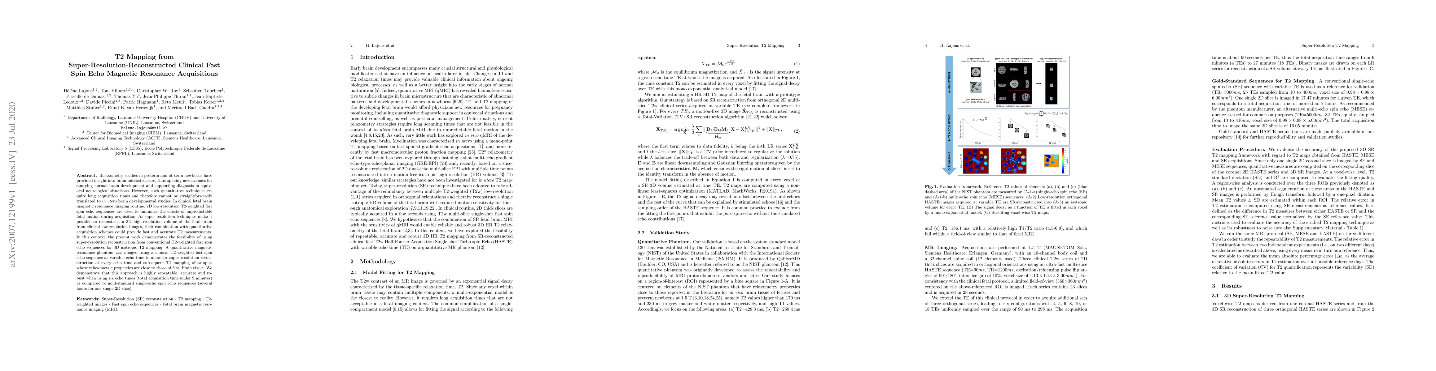
Discussion 0