Tailored Speckle Illumination Microscopy with Enhanced Sectioning and Image Quality
Publication
Metrics
Paper Preview
Abstract
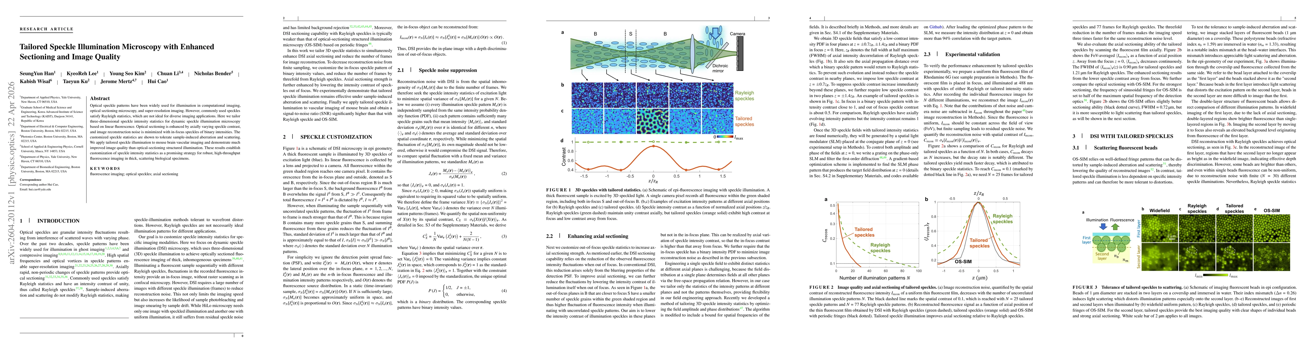
Optical speckle patterns have been widely used for illumination in computational imaging, optical sectioning microscopy, and super-resolution imaging. However, commonly used speckles satisfy Rayleigh statistics, which are not ideal for diverse imaging applications. Here we tailor three-dimensional speckle intensity statistics for dynamic speckle illumination microscopy based on linear fluorescence. Optical sectioning is enhanced by axially varying speckle contrast, and image reconstruction noise is minimized with in-focus speckles of binary intensities. The customized speckle statistics are shown to tolerate sample-induced aberration and scattering. We apply tailored speckle illumination to mouse brain vascular imaging and demonstrate much improved image quality than optical-sectioning structured illumination. These results establish customization of speckle intensity statistics as a promising strategy for robust, high-throughput fluorescence imaging in thick, scattering biological specimens.
AI Key Findings
Get AI-generated insights about this paper's methodology, results, significance, and more — seven facets brought into focus.
Discussion 0