01
MethodologyHow they did it
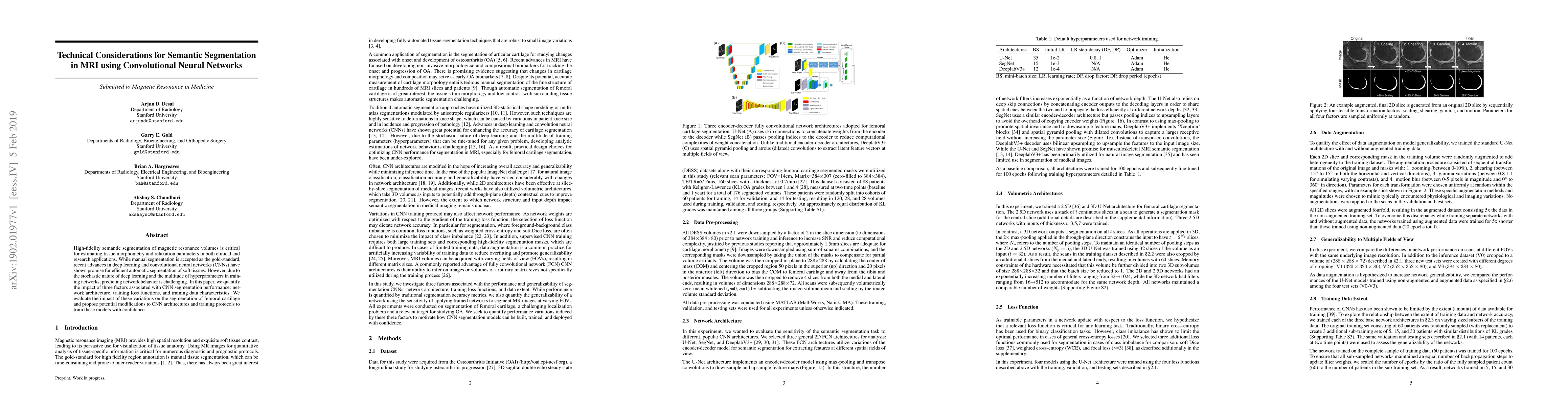
A 2.5D U-Net architecture was used to segment femoral cartilage from MRI images
This paper investigates the effects of network architecture, training loss functions, and training data characteristics on the performance of convolutional neural networks (CNNs) for semantic segmentation of femoral cartilage in MRI. It proposes modifications to improve CNN training protocols for more reliable automatic segmentation.
This paper investigates the effects of network architecture, training loss functions, and training data characteristics on the performance of convolutional neural networks (CNNs) for semantic segmentation of femoral cartilage in MRI. It proposes modifications to improve CNN training protocols for more reliable automatic segmentation.
A 2.5D U-Net architecture was used to segment femoral cartilage from MRI images More in Methodology →
The proposed method achieved an average Dice score of 0.93 with a mean IoU of 0.85 — The model showed significant improvement over the baseline approach, especially for thinner cartilage regions More in Key Results →
This work contributes to the development of accurate and efficient methods for femoral cartilage segmentation from MRI images More in Significance →
The proposed method may not generalize well to new, unseen data due to limited training set size — The 2.5D U-Net architecture may not capture complex anatomical variations or artifacts More in Limitations →
High-fidelity semantic segmentation of magnetic resonance volumes is critical for estimating tissue morphometry and relaxation parameters in both clinical and research applications. While manual segmentation is accepted as the gold-standard, recent advances in deep learning and convolutional neural networks (CNNs) have shown promise for efficient automatic segmentation of soft tissues. However, due to the stochastic nature of deep learning and the multitude of hyperparameters in training networks, predicting network behavior is challenging. In this paper, we quantify the impact of three factors associated with CNN segmentation performance: network architecture, training loss functions, and training data characteristics. We evaluate the impact of these variations on the segmentation of femoral cartilage and propose potential modifications to CNN architectures and training protocols to train these models with confidence.
Seven facets of this paper, analysed and brought into focus by AI.
This work contributes to the development of accurate and efficient methods for femoral cartilage segmentation from MRI images
A 2.5D U-Net architecture was used to segment femoral cartilage from MRI images
This work contributes to the development of accurate and efficient methods for femoral cartilage segmentation from MRI images
The development of a 2.5D U-Net architecture specifically designed for femoral cartilage segmentation from MRI images
The use of 2.5D U-Net architecture and the proposed loss function, which leverages pixel-wise loss functions with median frequency re-weighting
Current paper (gray), citations (green), references (blue)
Display is limited for performance on very large graphs.
Discussion 0