ArxivLens

Quick Summary:

This research proposes an improved technique for classifying meningioma tumours using a combination of texture measures, achieving a 92.50% accuracy. The method combines Gaussian Markov random field and run-length matrix texture features, outperforming individual texture measures and aiding pathologists in accurate diagnosis.

Journal Reference: Pattern Recognition, vol.43, pp.2043-2053, 2010
DOI: 10.1016/j.patcog.2010.01.005
arXiv Id: 1512.08049
Date Published: 2015-12-29
Date Updated: 2015-12-29

Categories:

Key Terms (TF-IDF):

texture (0.363)
combinations (0.243)
measures (0.238)
measures (0.224)
combinations (0.222)
accuracy (0.195)
features (0.186)
accuracy (0.176)
best (0.173)
features (0.172)
technique (0.172)
best (0.168)
colour (0.159)
discrimination (0.157)
optimum (0.157)
technique (0.156)
morphological (0.155)
segments (0.155)
classifier (0.154)
preparation (0.153)
License: http://arxiv.org/licenses/nonexclusive-distrib/1.0/

Summary:

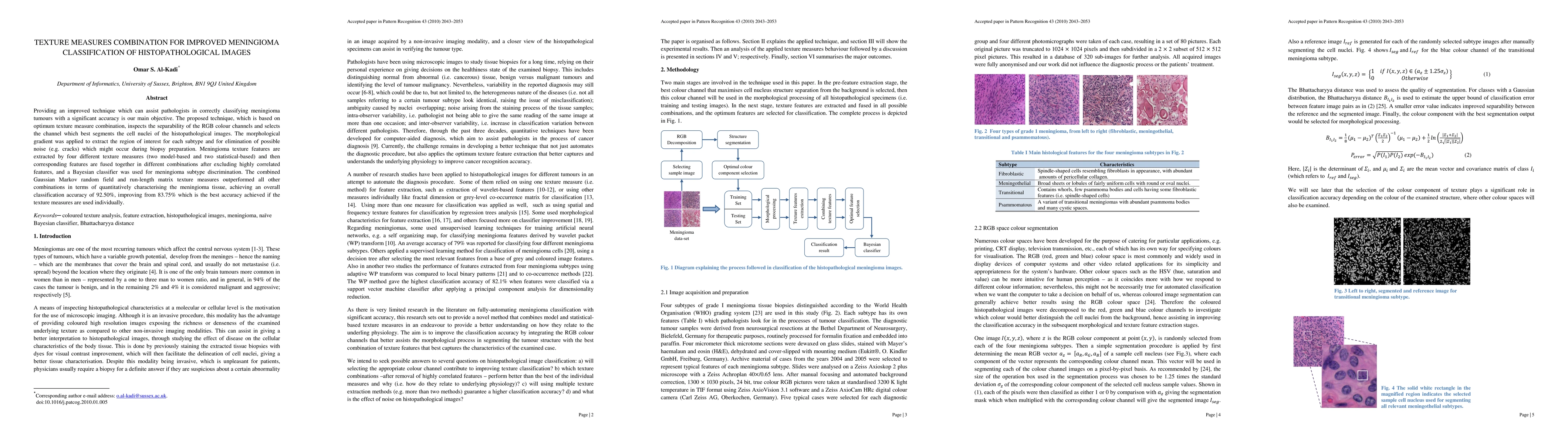
Providing an improved technique which can assist pathologists in correctly classifying meningioma tumours with a significant accuracy is our main objective. The proposed technique, which is based on optimum texture measure combination, inspects the separability of the RGB colour channels and selects the channel which best segments the cell nuclei of the histopathological images. The morphological gradient was applied to extract the region of interest for each subtype and for elimination of possible noise (e.g. cracks) which might occur during biopsy preparation. Meningioma texture features are extracted by four different texture measures (two model-based and two statistical-based) and then corresponding features are fused together in different combinations after excluding highly correlated features, and a Bayesian classifier was used for meningioma subtype discrimination. The combined Gaussian Markov random field and run-length matrix texture measures outperformed all other combinations in terms of quantitatively characterising the meningioma tissue, achieving an overall classification accuracy of 92.50%, improving from 83.75% which is the best accuracy achieved if the texture measures are used individually.

Citation & Reference Network Visualization

This graph shows how this paper is connected to other research. The current paper is shown in dark gray, papers that cite it are in green, and papers it references are in blue. You can:

Current Paper
Papers citing this work (Citations)
Papers cited by this work (References)

• Hover over any node to see the paper title

• Click a node to view that paper's details

• Drag nodes to rearrange the graph

• Use the zoom controls (bottom right) to zoom in/out

Citations and References

Title Authors Year Actions
Feed-forward networks using logistic regression and support vector machine for whole-slide breast cancer histopathology image classification
Arunadevi Karuppasamy, A. Abdesselam, R. Hedjam, Hamza M. Zidoum, Maiya Al-Bahri
2023
View Paper
Applications of discriminative and deep learning feature extraction methods for whole slide image analysis: A survey
Khaled A. Althelaya, Nauman Ullah Gila, M. Alzubaidi, Fahad Majeed, Marco Agus, J. Schneider, Mowafa J Househ
2023
View Paper
Early prediction of breast cancer based on the classification of HER‐2 and ER biomarkers using deep neural network
D. Saranyaraj, M. Manikandan
2023
View Paper
Classification of benign and malignant subtypes of breast cancer histopathology imaging using hybrid CNN-LSTM based transfer learning
Mahati Munikoti Srikantamurthy, V. S. Rallabandi, D. Dudekula, S. Natarajan, Junhyung Park
2023
View Paper
A deep learning framework identifies dimensional representations of Alzheimer’s Disease from brain structure
Zhijian Yang, I. Nasrallah, H. Shou, J. Wen, J. Doshi, M. Habes, G. Erus, A. Abdulkadir, S. Resnick, M. Albert, P. Maruff, J. Fripp, J. Morris, D. Wolk, C. Davatzikos
2021
View Paper
Artificial Intelligence Based Diagnostic Model for the Detection of Malaria Parasites from Microscopic Blood Images
G. Madhu, A. Govardhan
2021
View Paper
Diagnosis of skin cancer using machine learning techniques
A. Murugan, S. A. H. Nair, A. Preethi, K. P. S. Kumar
2021
View Paper
Colorectal cancer detection by immunofluorescence images of circulating tumor cells
Hsien-I Lin, Ying-Chih Chang
2021
View Paper
Objective Diagnosis for Histopathological Images Based on Machine Learning Techniques: Classical Approaches and New Trends
Naira Elazab, Hassan H. Soliman, Shaker El-Sappagh, S. Islam, Mohammed M Elmogy
2020
View Paper
Study of morphological and textural features for classification of oral squamous cell carcinoma by traditional machine learning techniques
Tabassum Yesmin Rahman, L. Mahanta, Hiten Choudhury, A. Das, J. Sarma
2020
View Paper
A Comprehensive Review of Markov Random Field and Conditional Random Field Approaches in Pathology Image Analysis
Yixin Li, Chen Li, Xiaoyan Li, Kai Wang, M. Rahaman, Changhao Sun, Hao Chen, Xinran Wu, Hong Zhang, Qian Wang
2020
View Paper
A Comprehensive Review for MRF and CRF Approaches in Pathology Image Analysis
Chen Li, Yixin Li, Changhao Sun, Hao Chen, Hong Zhang
2020
View Paper
Multi‐feature fusion of deep networks for mitosis segmentation in histological images
Yuan Zhang, Jin Chen, Xianzhu Pan
2020
View Paper
Automatic Bone Marrow Cellularity Estimation in H&E Stained Whole Slide Images
F. Nielsen, Mads Jozwiak Pedersen, Mathias Vassard Olsen, Morten Skaarup Larsen, R. Røge, A. Jørgensen
2019
View Paper
Mapping Digital Drainage Network Using Geoprocessing: A Case Study of Kali Gandaki River Basin, Nepal Himalaya
Feiyu Chen, B. Tian, B. Adhikari, Xiaoyun Gou
2019
View Paper
HIC-net: A deep convolutional neural network model for classification of histopathological breast images
Ş. Öztürk, B. Akdemir
2019
View Paper
AiCNNs (Artificially-integrated Convolutional Neural Networks) for Brain Tumor Prediction
Ansh Mittal, Deepika Kumar
2019
View Paper
Adaptive multidimensional fuzzy sets for texture modeling
P. Martínez-Jiménez, J. Chamorro-Martínez, J. Keller
2018
View Paper
Radiomics and machine learning may accurately predict the grade and histological subtype in meningiomas using conventional and diffusion tensor imaging
Y. Park, Jong-Kap Oh, S. You, Kyunghwa Han, S. Ahn, Y. Choi, Jong-Hee Chang, S. H. Kim, Seung-Koo Lee
2018
View Paper
Quantitative morphometric analysis of adult teleost fish by X-ray computed tomography
V. Weinhardt, Roman Shkarin, Tobias Wernet, J. Wittbrodt, T. Baumbach, F. Loosli
2018
View Paper
A novel computational method for automatic segmentation, quantification and comparative analysis of immunohistochemically labeled tissue sections
E. Casiraghi, V. Huber, M. Frasca, M. Cossa, Matteo Tozzi, L. Rivoltini, B. Leone, A. Villa, B. Vergani
2018
View Paper
Effective histopathological image area reduction method for real-time applications
Ş. Öztürk, B. Akdemir
2018
View Paper
Feature-based analysis of cell nuclei structure for classification of histopathological images
Pin Wang, Shan Xu, Yong-min Li, Lirui Wang, Q. Song
2018
View Paper
Randomized neural network based signature for color texture classification
Jarbas Joaci de Mesquita Sá Junior, A. Backes, O. Bruno
2018
View Paper
A HIERARCHICAL CLASSIFIER FOR MULTICLASS PROSTATE HISTOPATHOLOGY IMAGE GLEASON GRADING
Dheeb Albashish, S. Sahran, A. Abdullah, Mohammed Alweshah, Afzan Adam
2018
View Paper
Using wavelet sub-band and fuzzy 2-partition entropy to segment chronic lymphocytic leukemia images
T. A. Tosta, P. D. Faria, V. Batista, L. A. Neves, M. Z. Nascimento
2018
View Paper
LBP maps for improving fractal based texture classification
A. Backes, Jarbas Joaci de Mesquita Sá Junior
2017
View Paper
Randomized Neural Network Based Signature for Classification of Titanium Alloy Microstructures
Jarbas Joaci de Mesquita Sá Junior, A. Backes, O. Bruno
2017
View Paper
Feature extraction from histopathological images based on nucleus-guided convolutional neural network for breast lesion classification
Yushan Zheng, Zhi-guo Jiang, Feng-ying Xie, Haopeng Zhang, Yibing Ma, Huaqiang Shi, Yu Zhao
2017
View Paper
Texture analysis in liver of common carp (Cyprinus carpio) sub-chronically exposed to perfluorooctanoic acid
M. Manera, L. Giari, F. Vincenzi, C. Guerranti, J. Depasquale, G. Castaldelli
2017
View Paper
Using cell nuclei features to detect colon cancer tissue in hematoxylin and eosin stained slides
A. Jørgensen, Anders Munk Rasmussen, Niels K. Andersen, Simon Kragh Andersen, Jonas Emborg, R. Røge, L. Østergaard
2017
View Paper
Sparse Contribution Feature Selection and Classifiers Optimized by Concave-Convex Variation for HCC Image Recognition
Wenbo Pang, Huiyan Jiang, Siqi Li
2017
View Paper
Neuronal Texture Analysis in Murine Model of Down's Syndrome
A. Sarmiento, M. A. F. Granero, B. Galán, M. L. Montesinos, I. Fondón
2017
View Paper
Lumen-nuclei ensemble machine learning system for diagnosing prostate cancer in histopathology images
Dheeb Albashish, S. Sahran, A. Abdullah, Nordashima Abd Shukor, S. H. M. Pauzi
2017
View Paper
Multi-level learning approach for prostate histopathology images classification
S. Sahran, A. Abdullah, Dheeb Albashish, Sawsan Abu-Taleb
2017
View Paper
High-throughput fat quantifications of hematoxylin-eosin stained liver histopathological images based on pixel-wise clustering
Peng Shi, Junru Chen, Jinke Lin, Lurong Zhang
2017
View Paper
A Gabor Filter Texture Analysis Approach for Histopathological Brain Tumor Subtype Discrimination
Omar Sultan Al-Kadi
2017
View Paper
Nuclear spatial and spectral features based evolutionary method for meningioma subtypes classification in histopathology
Kiran Fatima, Hammad Majeed, H. Irshad
2017
View Paper
Texture analysis of parasitological liver fibrosis images
L. Moraru, Simona Moldovanu, A. Culea-Florescu, D. Bibicu, A. Ashour, N. Dey
2017
View Paper
Breast Cancer Detection using Support Vector Machine
Chithra Devi, S. D. Bharathi
2017
View Paper
An innovative neural network framework to classify blood vessels and tubules based on Haralick features evaluated in histological images of kidney biopsy
Vitoantonio Bevilacqua, Nicola Pietroleonardo, Vito Triggiani, Antonio Brunetti, A. D. Palma, M. Rossini, L. Gesualdo
2017
View Paper
DCE-MRI Texture Features for Early Prediction of Breast Cancer Therapy Response
Guillaume Thibault, A. Tudorica, Aneela Afzal, S. Chui, A. Naik, M. Troxell, K. Kemmer, K. Oh, N. Roy, Neda Jafarian, M. Holtorf, Wei Huang, Xubo B. Song
2017
View Paper
Synergies between texture features: an abstract feature based framework for meningioma subtypes classification
Hammad Majeed, Kiran Fatima
2017
View Paper
Ensemble Learning of Tissue Components for Prostate Histopathology Image Grading
Dheeb Albashish, S. Sahran, A. Abdullah, Nordashima Abd Shukor, S. H. M. Pauzi
2016
View Paper
Automated Quantitative Image Analysis of Hematoxylin-Eosin Staining Slides in Lymphoma Based on Hierarchical Kmeans Clustering
Peng Shi, Jing Zhong, Rongfang Huang, Jian-Jiao Lin
2016
View Paper
Fuzzy Statistical Matrices for Cell Classification
Guillaume Thibault, Izhak Shafran
2016
View Paper
Automated nuclei segmentation of malignant using level sets
A. Husham, Mohammed Hazim Alkawaz, T. Saba, A. Rehman, Jarallah Saleh Alghamdi
2016
View Paper
Multiscale Nakagami parametric imaging for improved liver tumor localization
Omar Sultan Al-Kadi
2016
View Paper
Quarter Plane ARMA Model for Analysis and Classification of Histopathology Images: Application to Cancer Detection
D. Vaishali, R. Ramesh, J. Christaline
2016
View Paper
Automated classification of brain tumor type in whole-slide digital pathology images using local representative tiles
Jocelyn Barker, A. Hoogi, A. Depeursinge, D. Rubin
2016
View Paper
Clinically-inspired automatic classification of ovarian carcinoma subtypes
A. Bentaieb, M. Nosrati, H. Li-Chang, D. Huntsman, G. Hamarneh
2016
View Paper
A Gravitational Model for Grayscale Texture Classification Applied to the pap-smear Database
Jarbas Joaci de Mesquita Sá Junior, A. Backes
2015
View Paper
Predicting Classifier Performance with Limited Training Data: Applications to Computer-Aided Diagnosis in Breast and Prostate Cancer
A. Basavanhally, Satish Viswanath, A. Madabhushi
2015
View Paper
Nuclear segmentation for skin cancer diagnosis from histopathological images
Pravda Jith Ray, S. Priya, T. Kumar
2015
View Paper
Identifying histological concepts on basal cell carcinoma images using nuclei based sampling and multi-scale descriptors
David Romo-Bucheli, Ricardo Moncayo, Angel Cruz-Roa, E. Romero
2015
View Paper
A Multiresolution Clinical Decision Support System Based on Fractal Model Design for Classification of Histological Brain Tumours
O. S. Al-Kadi
2015
View Paper
Analyse statistique de populations pour l'interprétation d'images histologiques. (Statistical analysis of populations for histological images interpretation)
M. Ali
2015
View Paper
Classification of Histological Images Based on the Stationary Wavelet Transform
M Z Nascimento, L. Neves, S C Duarte, Y. A. S. Duarte, V. R. Batista
2015
View Paper
A new texture and shape based technique for improving meningioma classification
Kiran Fatima, Arshia Arooj, Hammad Majeed
2014
View Paper
Automated classification of four types of developmental odontogenic cysts
A. Frydenlund, M. Eramian, T. Daley
2014
View Paper
Artificial crawler model for texture analysis on silk fibroin scaffolds
Bruno Brandoli Machado, W. Gonçalves, O. Bruno
2014
View Paper
A discriminant multi-scale histopathology descriptor using dictionary learning
D. Romo, Juan D. García-Arteaga, P. Arbeláez, E. Romero
2014
View Paper
Advanced Statistical Matrices for Texture Characterization: Application to Cell Classification
Guillaume Thibault, J. Angulo, F. Meyer
2014
View Paper
Automated Mitosis Detection in Color and Multi-spectral High-Content Images in Histopathology: Application to Breast Cancer Grading in Digital Pathology
H. Irshad
2014
View Paper
Computer-Aided Diagnosis of Skin Lesions Using Conventional Digital Photography: A Reliability and Feasibility Study
W. Chang, A. Huang, Chung-Yi Yang, Chien-Hung Lee, Yin‐Chun Chen, Tian-Yau Wu, Gwo-Shing Chen
2013
View Paper
Reviewing, selecting and evaluating features in distinguishing fine changes of global texture
Benhur Ortiz Jaramillo, Sergio A. Orjuela Vargas, L. Langenhove, G. Castellanos-Domínguez, W. Philips
2013
View Paper
Exhaustive comparison of colour texture features and classification methods to discriminate cells categories in histological images of fish ovary
Encarnación González-Rufino, P. Carrión, E. Cernadas, M. Delgado, R. Domínguez-Petit
2013
View Paper
Multi-channels statistical and morphological features based mitosis detection in breast cancer histopathology
H. Irshad, L. Roux, Daniel Racoceanu
2013
View Paper
Practical quantification of necrosis in histological whole-slide images
A. Homeyer, A. Schenk, J. Arlt, U. Dahmen, O. Dirsch, H. Hahn
2013
View Paper
Meningioma subtype classification using morphology features and random forests
H. Strange, R. Zwiggelaar
2013
View Paper
Predicting Future Morphological Changes of Lesions from Radiotracer Uptake in 18F-FDG-PET Images
Ulas Bagci, Jianhua Yao, Kirsten Miller-Jaster, Xinjian Chen, D. Mollura
2013
View Paper
Automated mitosis detection in histopathology using morphological and multi-channel statistics features
H. Irshad
2013
View Paper
Automated mitosis detection using texture, SIFT features and HMAX biologically inspired approach
H. Irshad, Sepehr Jalali, L. Roux, Daniel Racoceanu, Lim Joo Hwee, G. Naour, F. Capron
2013
View Paper
HISTOPATHOLOGICAL IMAGE ANALYSIS USING IMAGE PROCESSING TECHNIQUES : AN OVERVIEW
A. Belsare, M. Mushrif
2012
View Paper
Micro‐structural tissue analysis for automatic histopathological image annotation
Gloria Díaz, E. Romero
2012
View Paper
Automated oral cancer identification using histopathological images: a hybrid feature extraction paradigm.
M. M. R. Krishnan, Vikram Venkatraghavan, U. R. Acharya, M. Pal, R. Paul, L. C. Min, A. Ray, J. Chatterjee, C. Chakraborty
2012
View Paper
Automated Diagnosis of Oral Cancer Using Higher Order Spectra Features and Local Binary Pattern: A Comparative Study
M. M. R. Krishnan, U. R. Acharya, C. Chakraborty, A. Ray
2011
View Paper
Textural characterization of histopathological images for oral sub-mucous fibrosis detection.
M. M. R. Krishnan, P. Shah, A. Choudhary, C. Chakraborty, R. Paul, A. Ray
2011
View Paper
Optimizing automated characterization of liver fibrosis histological images by investigating color spaces at different resolutions
D. Mahmoud-Ghoneim
2011
View Paper
Texture analysis of liver fibrosis microscopic images: a study on the effect of biomarkers.
A. Amin, D. Mahmoud-Ghoneim
2011
View Paper
Meningioma subtype classification: A survey
Kiran Fatima, Hammad Majeed
2010
View Paper
A study on the clinical classification model for the hand venule of baby pneumonia based on decision tree algorithm
Yuelin Lu, Benzheng Wei, Xinjun Wang
2010
View Paper
Comparative analysis of spatial and transform domain methods for meningioma subtype classification
H. Qureshi, N. Rajpoot
2010
View Paper
File A deep learning framework identifies dimensional representations of Alzheimer ’ s Disease from brain structure
2021
View Paper
GLCM and GLRLM Based Texture Analysis: Application to Brain Cancer Diagnosis Using Histopathology Images
Vaishali Durgamahanthi, J. Christaline, A. S. Edward
2020
View Paper
Classification of Medical Images using Artificial Neural Network
Akash Verma
2020
View Paper
Automated analysis of necrosis and steatosis in histological images : Practical solutions for coping with heterogeneity and variability (Automatisierte Analyse von Nekrose und Steatose in histologischen Bildern)
A. Homeyer
2019
View Paper
Automatic Detection of Prostate Cancer by Computer Aided Diagnosis
S. Gurav
2017
View Paper
A Two Phase Hybrid Classifier based on Structure Similarities and Textural Features for Accurate Meningioma Classification
Kiran Fatima, Hammad Majeed
2017
View Paper
Fractals for Biomedical Texture Analysis
O. S. Al-Kadi
2017
View Paper
Texture-Based Evolutionary Method for Cancer Classification in Histopathology
Kiran Fatima, Hammad Majeed
2017
View Paper
Triaging Diagnostically Relevant Regions from Pathology Whole Slides of Breast Cancer: A Texture Based Approach
Mohammad Peikari, M. Gangeh, J. Zubovits, G. Clarke, Anne L. Martel
2016
View Paper
Computer aided classification of histopathological damage in images of haematoxylin and eosin stained human skin
J. Haggerty
2016
View Paper
Computational Fractal-Based Analysis of Brain Tumor Microvascular Networks
A. Ieva, O. S. Al-Kadi, Omar Sultan Al-Kadi
2016
View Paper
Histological Fractal-Based Classification of Brain Tumors
O. S. Al-Kadi, O. S. Al-Kadi, A. Ieva
2016
View Paper
Methods for Nuclei Detection, Segmentation, and Classification in Digital Histopathology: A Review—Current Status and Future Potential
H. Irshad, A. Veillard, L. Roux, Daniel Racoceanu
2014
View Paper
Image Analysis Methods and Tools for Digital Histopathology Applications Relevant to Breast Cancer Diagnosis
Andreas Kårsnäs
2014
View Paper
Adaptive Gray Level Co-occurrence Probabilities for Texture Segmentation Based on Coarse-to-fine Strategy
Yao-Hung Hubert Tsai
2014
View Paper
Computer-aided colorectal tumor classification in NBI endoscopy using local features
Toru Tamaki, Junki Yoshimuta, Misato Kawakami, B. Raytchev, K. Kaneda, S. Yoshida, Yoshito Takemura, K. Onji, R. Miyaki, Shinji Tanaka
2013
View Paper
Higher order statistical analysis of nanoscale resolution images of unstained tissue sections for cancer detection
Alexandru Foias, E. Perrin, V. Vrabie
2013
View Paper
İÇERİK TABANLI MEDİKAL İMGE ERİŞİMİNDE GABOR DOKUSAL ÖZNİTELİKLERİNİN VERİMLİLİĞİ
Muhammet Baykara, Burhan Ergen
2011
View Paper
Tumour Grading and Discrimination based on Class Assignment and Quantitative Texture Analysis Techni
O. S. Al-Kadi
2009
View Paper

Comments (0)

This website uses cookies to ensure you get the best experience. Learn more