ArxivLens

Quick Summary:

This paper proposes a 2-Stage Training (2ST-UNet) system using a ResNet34 backbone for segmenting pneumothorax in chest X-rays. The method employs a lower resolution training followed by higher resolution retraining, along with Stochastic Weight Averaging and data augmentation. The model ranks 124 out of 1,475 with a mean Dice Similarity Coefficient of 0.8356.

arXiv Id: 2009.02805
Date Published: 2020-09-09
Date Updated: 2020-09-09

Categories:

Key Terms (TF-IDF):

augmentation (0.223)
augmentation (0.203)
training (0.192)
training (0.190)
images (0.183)
stage (0.182)
stage (0.180)
images (0.177)
identified (0.162)
identified (0.159)
trained (0.155)
network (0.152)
trained (0.151)
network (0.147)
dataset (0.147)
resolution (0.146)
dataset (0.143)
ray (0.143)
resolution (0.141)
ray (0.140)
License: http://arxiv.org/licenses/nonexclusive-distrib/1.0/

Summary:

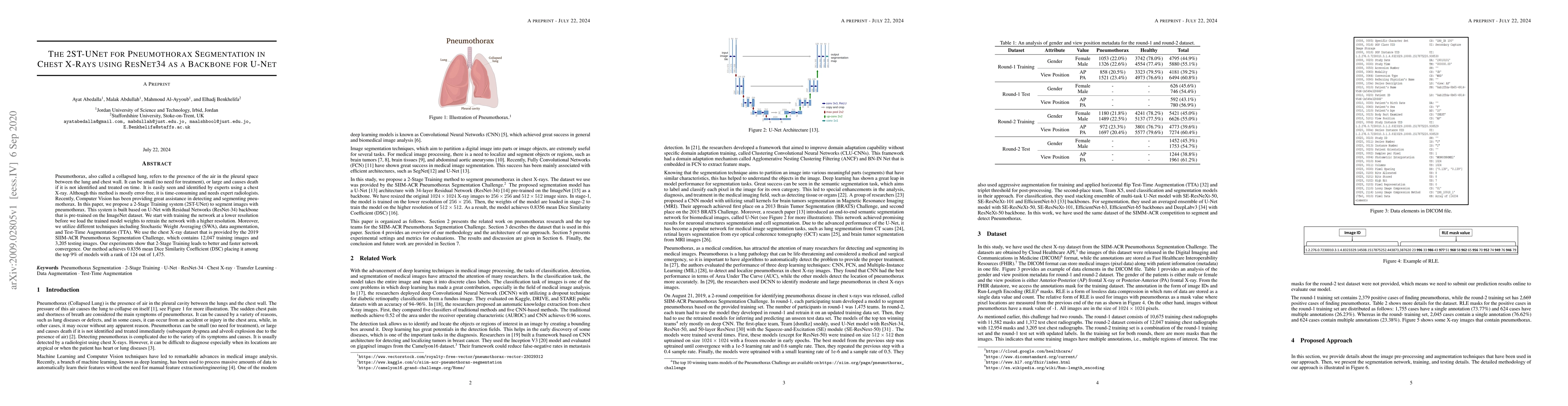
Pneumothorax, also called a collapsed lung, refers to the presence of the air in the pleural space between the lung and chest wall. It can be small (no need for treatment), or large and causes death if it is not identified and treated on time. It is easily seen and identified by experts using a chest X-ray. Although this method is mostly error-free, it is time-consuming and needs expert radiologists. Recently, Computer Vision has been providing great assistance in detecting and segmenting pneumothorax. In this paper, we propose a 2-Stage Training system (2ST-UNet) to segment images with pneumothorax. This system is built based on U-Net with Residual Networks (ResNet-34) backbone that is pre-trained on the ImageNet dataset. We start with training the network at a lower resolution before we load the trained model weights to retrain the network with a higher resolution. Moreover, we utilize different techniques including Stochastic Weight Averaging (SWA), data augmentation, and Test-Time Augmentation (TTA). We use the chest X-ray dataset that is provided by the 2019 SIIM-ACR Pneumothorax Segmentation Challenge, which contains 12,047 training images and 3,205 testing images. Our experiments show that 2-Stage Training leads to better and faster network convergence. Our method achieves 0.8356 mean Dice Similarity Coefficient (DSC) placing it among the top 9% of models with a rank of 124 out of 1,475.

Citation & Reference Network Visualization

This graph shows how this paper is connected to other research. The current paper is shown in dark gray, papers that cite it are in green, and papers it references are in blue. You can:

Current Paper
Papers citing this work (Citations)
Papers cited by this work (References)

• Hover over any node to see the paper title

• Click a node to view that paper's details

• Drag nodes to rearrange the graph

• Use the zoom controls (bottom right) to zoom in/out

Citations and References

Title Authors Year Actions
Enhancing Medical Image Analysis with CNN and MobileNet: A Particle Swarm Optimization Approach
Asmaa Ghali Sabea1, Maryam Jawad Kadhim2, Ali Fahem, Mohammed Ibrahim Mahdi4
2025
View Paper
Integrating deep learning and machine learning for improved CKD-related cortical bone assessment in HRpQCT images: A pilot study
Youngjun Lee, Wikum R. Bandara, Sangjun Park, Miran Lee, Choongboem Seo, Sunwoo Yang, Kenneth J. Lim, Sharon M. Moe, Stuart J. Warden, Rachel K. Surowiec
2024
View Paper
A Super-Resolution Framework for Emotion Recognition Based on EEG Signals
Guofa Li, Yufei Yuan, Chuzhao Li, Qingkun Li, Wenbo Li, Zhenning Li, Paul Green
2024
View Paper
Comprehensive Experiments on Breast Cancer Hematoxylin and Eosin-stained Images Using UNet
Emily Jackson, Faye Le, Je'Dae I. Lisbon, Max Coleman, Jordyn Burman, Astrid Wonderley, Sepehr Eshaghian, Sanghoon Lee
2024
View Paper
Automatic Segmentation of Bone Marrow Lesions on MRI Using a Deep Learning Method
Raj Ponnusamy, Ming Zhang, Yue Wang, Xinyue Sun, Mohammad Chowdhury, J. Driban, T. McAlindon, Juan Shan
2024
View Paper
Multi-class Tissue Segmentation of CT images using an Ensemble Deep Learning method
N. Mahmoodian, Sumit Chakrabarty, Marilena Georgiades, Maciej Pech, C. Hoeschen
2023
View Paper
Validation of automatically measured T1 map cortico-medullary difference (ΔT1) for eGFR and fibrosis assessment in allograft kidneys
Ibtisam Aslam, Fariha Aamir, Miklós Kassai, L. Crowe, P. Poletti, S. Seigneux, S. Moll, Lena Berchtold, J. Vallée
2023
View Paper
Developing a Tuned Three-Layer Perceptron Fed with Trained Deep Convolutional Neural Networks for Cervical Cancer Diagnosis
Shervan Fekri-Ershad, M. Alsaffar
2023
View Paper
CNN based Intelligent Streetlight Management Using Smart CCTV Camera and Semantic Segmentation
Md Sakib Ullah Sourav, Huidong Wang, Mohammad Raziuddin Chowdhury, R. Sulaiman
2022
View Paper
Deep Learning-Based Computer-Aided Pneumothorax Detection Using Chest X-ray Images
Priyanka Malhotra, Sheifali Gupta, Deepika Koundal, Atef Zaguia, Manjit Kaur, Heung-No Lee
2022
View Paper
PTXNet: An extended UNet model based segmentation of pneumothorax from chest radiography images
Aarya Patel, Ankit Vidyarthi
2021
View Paper
Energy Efficient Automatic Streetlight Controlling System using Semantic Segmentation
Md Sakib Ullah Sourav, Huidong Wang
2022
View Paper

Comments (0)

This website uses cookies to ensure you get the best experience. Learn more