The Deep Poincar\'e Map: A Novel Approach for Left Ventricle Segmentation
Publication
Metrics
AI Quick Summary
A new method for segmenting the left ventricle in cardiac MRI images uses a deep learning model to guide an agent through the image, tracing out its boundary using the Poincaré map as a stopping criterion. The approach outperforms previous methods on multiple metrics.
Paper Preview
Abstract
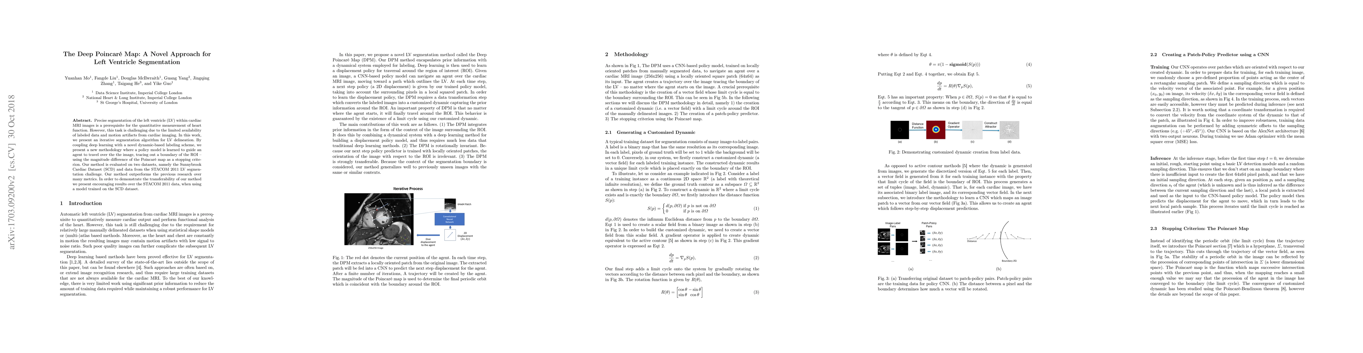
Precise segmentation of the left ventricle (LV) within cardiac MRI images is a prerequisite for the quantitative measurement of heart function. However, this task is challenging due to the limited availability of labeled data and motion artifacts from cardiac imaging. In this work, we present an iterative segmentation algorithm for LV delineation. By coupling deep learning with a novel dynamic-based labeling scheme, we present a new methodology where a policy model is learned to guide an agent to travel over the the image, tracing out a boundary of the ROI -- using the magnitude difference of the Poincar\'e map as a stopping criterion. Our method is evaluated on two datasets, namely the Sunnybrook Cardiac Dataset (SCD) and data from the STACOM 2011 LV segmentation challenge. Our method outperforms the previous research over many metrics. In order to demonstrate the transferability of our method we present encouraging results over the STACOM 2011 data, when using a model trained on the SCD dataset.
AI Key Findings
Get AI-generated insights about this paper's methodology, results, significance, and more — seven facets brought into focus.
Impact
Paper Details
PDF Preview
Key Terms
Citation Network
Current paper (gray), citations (green), references (blue)
Display is limited for performance on very large graphs.
Discussion 0