The Detection of Nanoscale Membrane Bending with Polarized Localization Microscopy
Publication
Metrics
AI Quick Summary
This paper introduces Polarized Localization Microscopy (PLM), a super-resolution optical imaging technique combining polarized total internal reflection fluorescence microscopy (TIRFM) and single-molecule localization microscopy to detect nanoscale membrane curvature and correlate it with single-molecule dynamics. PLM offers a 10x faster and more sensitive method for membrane curvature detection compared to 3D single-molecule localization techniques.
Paper Preview
Abstract
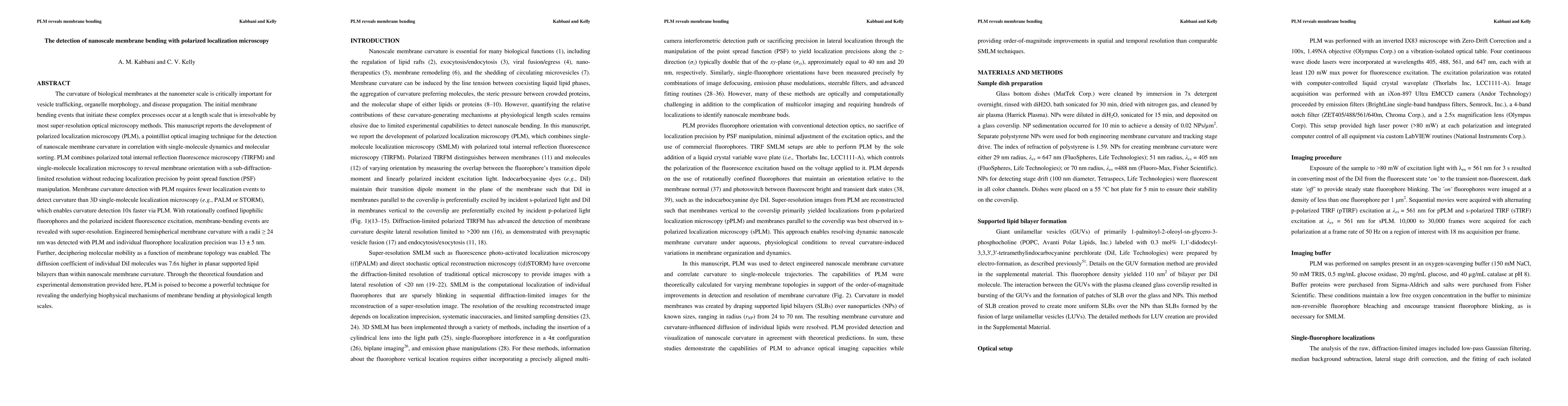
The curvature of biological membranes at the nanometer scale is critically important for vesicle trafficking, organelle morphology, and disease propagation. This manuscript reports the development of Polarized Localization Microscopy (PLM), a pointillist optical imaging technique for the detection of nanoscale membrane curvature in correlation with single-molecule dynamics and molecular sorting. PLM combines polarized total internal reflection fluorescence microscopy (TIRFM) and single-molecule localization microscopy to reveal membrane orientation with sub-diffraction-limited resolution without reducing localization precision by point spread function (PSF) manipulation. Further, membrane curvature detection with PLM requires fewer localization events to detect curvature than 3D single-molecule localization microscopy (e.g., PALM or STORM), which enables curvature detection 10x faster via PLM than via 3D single-molecule localizations. With high sensitivity, PLM detects curvature with provides super resolution images with >10x signal-to-noise enhancement from diffraction-limited polarized TIRFM. With rotationally confined lipophilic fluorophores and the polarized incident fluorescence excitation, membrane-bending events are revealed with super-resolution. Engineered hemispherical membrane curvature with a radius >= 24 nm was detected with PLM with individual fluorophore have a localization precision of 13 +/- 5 nm. Further, deciphering molecular dynamics as a function of membrane topology was enabled. The diffusion coefficient of individual DiI molecules was 7.6x higher in planar supported lipid bilayers than within nanoscale membrane curvature.
AI Key Findings
Get AI-generated insights about this paper's methodology, results, significance, and more — seven facets brought into focus.
Impact
Paper Details
PDF Preview
Key Terms
Citation Network
Current paper (gray), citations (green), references (blue)
Display is limited for performance on very large graphs.
Discussion 0