Publication
Metrics
AI Quick Summary
The paper presents the IDEAS open-source MRI dataset comprising preprocessed scans from 442 epilepsy patients who underwent neurosurgery, aimed at enhancing machine learning in detecting cerebral abnormalities. The dataset includes detailed demographic and clinical information, and its release is expected to advance computational methods in neurology.
Paper Preview
Abstract
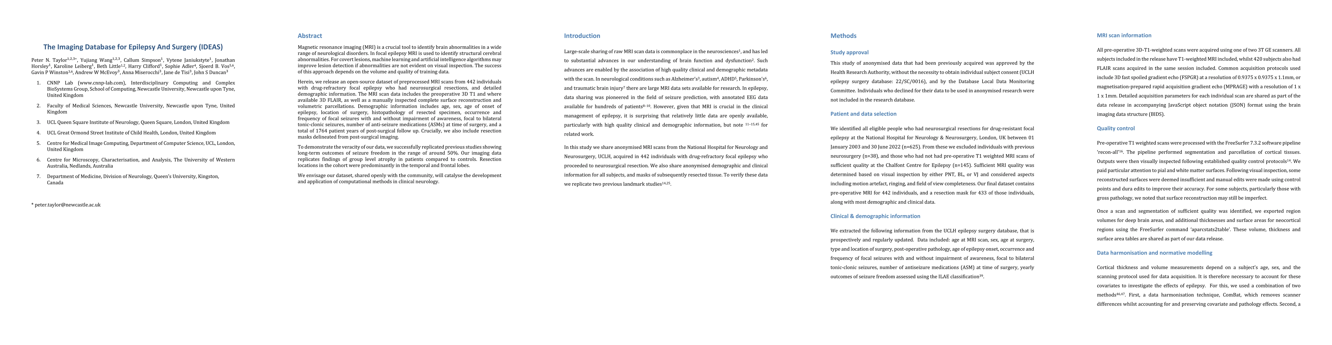
Magnetic resonance imaging (MRI) is a crucial tool to identify brain abnormalities in a wide range of neurological disorders. In focal epilepsy MRI is used to identify structural cerebral abnormalities. For covert lesions, machine learning and artificial intelligence algorithms may improve lesion detection if abnormalities are not evident on visual inspection. The success of this approach depends on the volume and quality of training data. Herein, we release an open-source dataset of preprocessed MRI scans from 442 individuals with drug-refractory focal epilepsy who had neurosurgical resections, and detailed demographic information. The MRI scan data includes the preoperative 3D T1 and where available 3D FLAIR, as well as a manually inspected complete surface reconstruction and volumetric parcellations. Demographic information includes age, sex, age of onset of epilepsy, location of surgery, histopathology of resected specimen, occurrence and frequency of focal seizures with and without impairment of awareness, focal to bilateral tonic-clonic seizures, number of anti-seizure medications (ASMs) at time of surgery, and a total of 1764 patient years of post-surgical follow up. Crucially, we also include resection masks delineated from post-surgical imaging. To demonstrate the veracity of our data, we successfully replicated previous studies showing long-term outcomes of seizure freedom in the range of around 50%. Our imaging data replicates findings of group level atrophy in patients compared to controls. Resection locations in the cohort were predominantly in the temporal and frontal lobes. We envisage our dataset, shared openly with the community, will catalyse the development and application of computational methods in clinical neurology.
AI Key Findings
Get AI-generated insights about this paper's methodology, results, significance, and more — seven facets brought into focus.
Impact
Paper Details
Authors
PDF Preview
Key Terms
Citation Network
Current paper (gray), citations (green), references (blue)
Display is limited for performance on very large graphs.
Discussion 0