01
MethodologyHow they did it
Brief description of the research methodology used
This study investigates the morphology of cell spheroids under simple shear flow using a parallel plate apparatus, revealing that spheroids rotate and lose size over time due to outer cell detachment, influenced by the shear stress. The findings provide a method to quantify flow-induced effects on cell-cell adhesion, relevant to mechanobiology.
This study investigates the morphology of cell spheroids under simple shear flow using a parallel plate apparatus, revealing that spheroids rotate and lose size over time due to outer cell detachment, influenced by the shear stress. The findings provide a method to quantify flow-induced effects on cell-cell adhesion, relevant to mechanobiology.
Brief description of the research methodology used More in Methodology →
Main finding 1 — Main finding 2 More in Key Results →
Why this research is important and its potential impact More in Significance →
Limitation 1 — Limitation 2 More in Limitations →
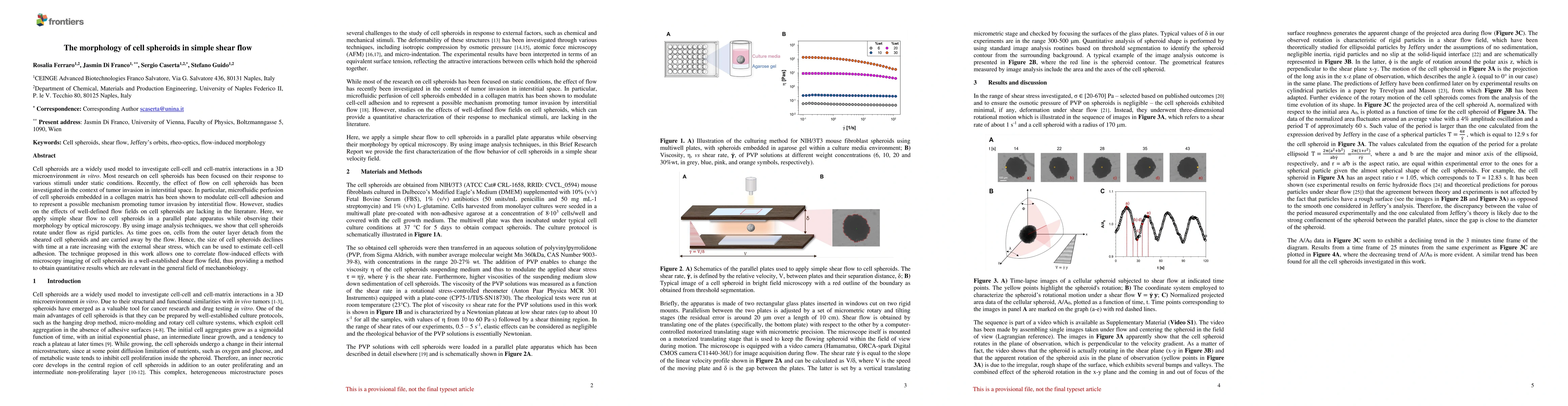
Cell spheroids are a widely used model to investigate cell-cell and cell-matrix interactions in a 3D microenvironment in vitro. Most research on cell spheroids has been focused on their response to various stimuli under static conditions. Recently, the effect of flow on cell spheroids has been investigated in the context of tumor invasion in interstitial space. In particular, microfluidic perfusion of cell spheroids embedded in a collagen matrix has been shown to modulate cell-cell adhesion and to represent a possible mechanism promoting tumor invasion by interstitial flow. However, studies on the effects of well-defined flow fields on cell spheroids are lacking in the literature. Here, we apply simple shear flow to cell spheroids in a parallel plate apparatus while observing their morphology by optical microscopy. By using image analysis techniques, we show that cell spheroids rotate under flow as rigid particles. As time goes on, cells from the outer layer detach from the sheared cell spheroids and are carried away by the flow. Hence, the size of cell spheroids declines with time at a rate increasing with the external shear stress, which can be used to estimate cell-cell adhesion. The technique proposed in this work allows one to correlate flow-induced effects with microscopy imaging of cell spheroids in a well-established shear flow field, thus providing a method to obtain quantitative results which are relevant in the general field of mechanobiology.
Seven facets of this paper, analysed and brought into focus by AI.
Why this research is important and its potential impact
Brief description of the research methodology used
Why this research is important and its potential impact
Main technical or theoretical contribution
What makes this work novel or different from existing research
Current paper (gray), citations (green), references (blue)
Display is limited for performance on very large graphs.
Discussion 0