This paper introduces a novel benchmark dataset, RETA, for retinal vascular tree analysis, featuring 81 labeled vessel masks, aiming to improve automated vessel segmentation and analysis. The dataset includes detailed annotations and is validated for superior quality, with the annotation software made publicly available for further research.
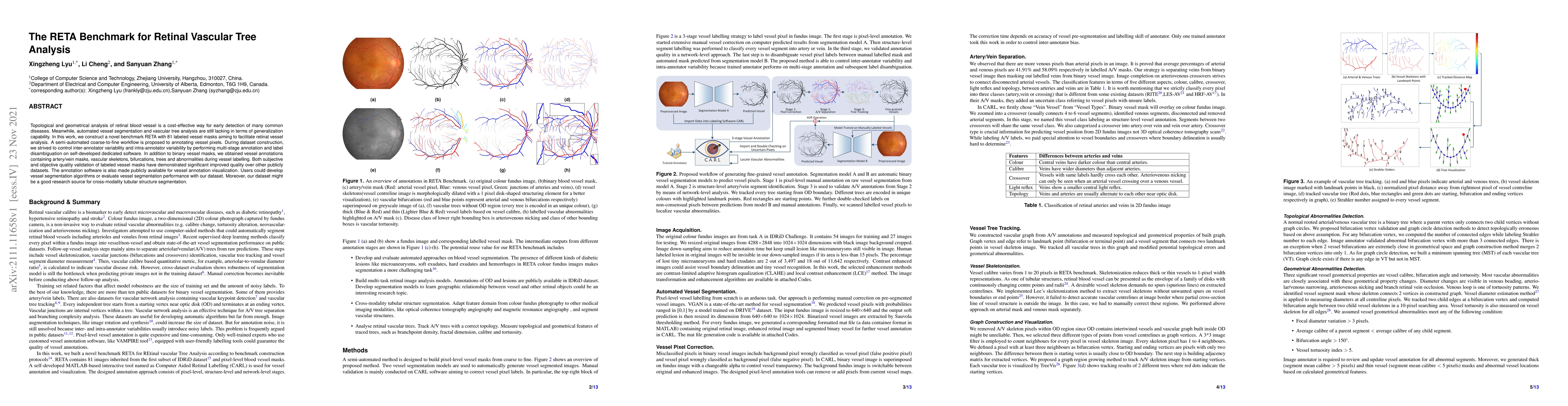
Topological and geometrical analysis of retinal blood vessel is a cost-effective way for early detection of many common diseases. Meanwhile, automated vessel segmentation and vascular tree analysis are still lacking in terms of generalization capability. In this work, we construct a novel benchmark RETA with 81 labeled vessel masks aiming to facilitate retinal vessel analysis. A semi-automated coarse-to-fine workflow is proposed to annotating vessel pixels. During dataset construction, we strived to control inter-annotator variability and intra-annotator variability by performing multi-stage annotation and label disambiguation on self-developed dedicated software. In addition to binary vessel masks, we obtained vessel annotations containing artery/vein masks, vascular skeletons, bifurcations, trees and abnormalities during vessel labelling. Both subjective and objective quality validation of labeled vessel masks have demonstrated significant improved quality over other publicly datasets. The annotation software is also made publicly available for vessel annotation visualization. Users could develop vessel segmentation algorithms or evaluate vessel segmentation performance with our dataset. Moreover, our dataset might be a good research source for cross-modality tubular structure segmentation.
Get AI-generated insights about this paper's methodology, results, and significance.
Current paper (gray), citations (green), references (blue)
Li Li, Junjie Yang, Zhihao Zhao et al.
Title | Authors | Year | Actions |
---|---|---|---|
Exploring a multi-path U-net with probability distribution attention and cascade dilated convolution for precise retinal vessel segmentation in fundus images
|
Ruihong Zhang, Guosong Jiang
|
2025
|
View Paper |
Evaluating Tree-Shaped Network Similarity via Morphological Feature Matching With Applications
|
Mona A. Ahmed, Omar M. Salim, M. Hassan, H. Dorrah, W. Gabr
|
2024
|
View Paper |
VascX Models: Model Ensembles for Retinal Vascular Analysis from Color Fundus Images
|
Jose Vargas Quiros, Bart Liefers, Karin A. van Garderen, Jeroen Vermeulen, Eyened Reading Center, Sinergia Consortium, Caroline Klaver
|
2024
|
View Paper |
Deep learning based approach: automated gingival inflammation grading model using gingival removal strategy
|
Chang Wen, Xueying Bai, Jiaxin Yang, Sihong Li, Xiaoxuan Wang, Dong Yang
|
2024
|
View Paper |
3D Vessel Graph Generation Using Denoising Diffusion
|
Chinmay Prabhakar, Suprosanna Shit, Fabio Musio, Kaiyuan Yang, Tamaz Amiranashvili, J. Paetzold, H. Li, Bjoern H Menze
|
2024
|
View Paper |
Task-Aware Transformer For Partially Supervised Retinal Fundus Image Segmentation
|
Hailong Zeng, Jianfeng Liu, Yixiong Liang
|
2024
|
View Paper |
SSL-ENSAM: Utilizing Sam for Semi-Supervised Retinal Vessel Segmentation with Quality-Aware Enhancement
|
Mengxian He, Minqing Zhang, Hing Lam Chang, Xiaoyi Feng, Mengdi Gao, Shuai Jiang, Wu Yuan
|
2024
|
View Paper |
Many birds, one stone: Medical image segmentation with multiple partially labeled datasets
|
Qing Liu, Hailong Zeng, Zhaodong Sun, Xiaobai Li, Guoying Zhao, Yixiong Liang
|
2024
|
View Paper |
FISBe: A Real-World Benchmark Dataset for Instance Segmentation of Long-Range thin Filamentous Structures
|
Lisa Mais, Peter Hirsch, Claire Managan, Ramya Kandarpa, J. L. Rumberger, Annika Reinke, Lena Maier-Hein, Gudrun Ihrke, Dagmar Kainmueller
|
2024
|
View Paper |
MAPLES-DR: MESSIDOR Anatomical and Pathological Labels for Explainable Screening of Diabetic Retinopathy
|
Gabriel Lepetit-Aimon, Clément Playout, M. Boucher, R. Duval, Michael H. Brent, Farida Cheriet
|
2024
|
View Paper |
Multi-site, Multi-domain Airway Tree Modeling
|
Minghui Zhang, Yang Wu, Hanxiao Zhang, Yulei Qin, Hao Zheng, Wen Tang, Corey W. Arnold, Chenhao Pei, Peng Yu, Yang Nan, Guangyao Yang, Simon L. F. Walsh, D. Marshall, M. Komorowski, Puyang Wang, Dazhou Guo, Dakai Jin, Yanan Wu, Shuiqing Zhao, Runsheng Chang, Boyu Zhang, Xing Lu, Abdul Qayyum, Moona Mazher, Qi Su, Yonghuang Wu, Ying'ao Liu, Yufei Zhu, Jiancheng Yang, A. Pakzad, B. Rangelov, R. Estépar, C. Cano-Espinosa, Jiayuan Sun, Guangyao Yang, Yun Gu
|
2023
|
View Paper |
Enhancing Vessel Segment Extraction in Retinal Fundus Images Using Retinal Image Analysis and Six Sigma Process Capability Index
|
S. Badawi, Maen Takruri, I. Elbadawi, I. Chaudhry, N. Mahar, A. Nileshwar, Emad Mosalam
|
2023
|
View Paper |
Generation of Structurally Realistic Retinal Fundus Images with Diffusion Models
|
Sojung Go, Younghoon Ji, Sang Jun Park, Soochahn Lee
|
2023
|
View Paper |
MSA-UNet: Retinal vessel segmentation method based on multiple spatial attention mechanism
|
Junhua Geng, Jinping Li, Xiao Wang
|
2023
|
View Paper |
Large scale crowdsourced radiotherapy segmentations across a variety of cancer anatomic sites
|
K. Wahid, D. Lin, B. Nelms, R. He, M. Naser, S. Duke, M. Sherer, J. Christodouleas, A. Mohamed, M. Cislo, J. Murphey, C. Fuller, E. Gillespie
|
2023
|
View Paper |
Large-scale crowdsourced radiotherapy segmentations across a variety of cancer anatomic sites: Interobserver expert/non-expert and multi-observer composite tumor and normal tissue delineation annotation from a prospective educational challenge
|
K. Wahid, Diana Lin, Onur Sahin, M. Cislo, Benjamin E. Nelms, R. He, M. Naser, Simon Duke, M. Sherer, J. Christodouleas, A. Mohamed, James D Murphy, C. Fuller, Erin F. Gillespie
|
2022
|
View Paper |
Fractal dimension of retinal vasculature as an image quality metric for automated fundus image analysis systems
|
Xingzheng Lyu, Purvish Jajal, Muhammad Zeeshan Tahir, S. Zhang
|
2022
|
View Paper |
Medical Image Analysis
|
Minghui Zhang, Yang Wu, Hanxiao Zhang, Yulei Qin, Hao Zheng, Wen Tang, C. Arnold, Chenhao Pei, Pengxin Yu, Yang Nan, Guangyao Yang, Simon L. F. Walsh, D. C. Marshall, M. Komorowski, Puyang Wang, Dazhou Guo, D. Jin, Yanan Wu, Shuiqing Zhao, Runsheng Chang, Boyu Zhang, Xingxin Lu, Abdul Qayyum, Moona Mazher, Qinzhi Su, Yonghuang Wu, Ying'ao Liu, Yufei Zhu, Jiancheng Yang, A. Pakzad, Bojidar, Rangelov, Raúl San José Estépar, C. Espinosa, Jiayuan Sun, Guangyao Yang, Yun Gu
|
2023
|
View Paper |
Medical Image Analysis
|
Minghui Zhang, Yang Wu, Hanxiao Zhang, Yulei Qin, Hao Zheng, Wen Tang, C. Arnold, Chenhao Pei, Pengxin Yu, Yang Nan, Guangyao Yang, Simon L. F. Walsh, D. C. Marshall, M. Komorowski, Puyang Wang, Dazhou Guo, D. Jin, Yanan Wu, Shuiqing Zhao, Runsheng Chang, Boyu Zhang, Xing-Pu Lv, Abdul Qayyum, Moona Mazher, Qinzhi Su, Yonghuang Wu, Ying'ao Liu, Yufei Zhu, Jiancheng Yang, A. Pakzad, B. Rangelov, Raúl San José Estépar, C. Espinosa, Jiayuan Sun, Guangyao Yang, Yun Gu
|
View Paper |
Comments (0)