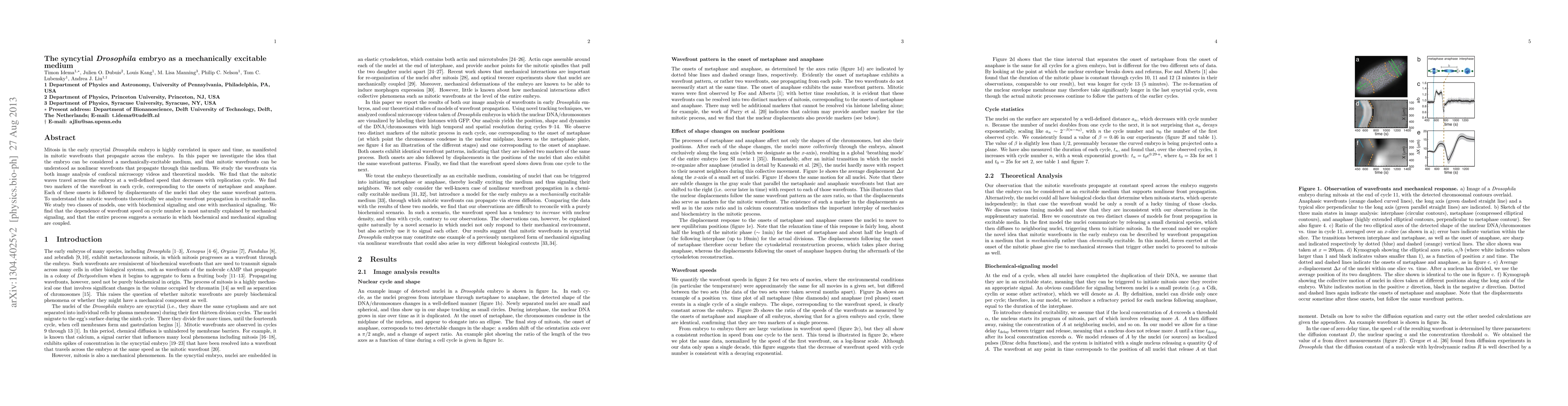
Publication
Metrics
AI Quick Summary
This paper explores the early syncytial Drosophila embryo as a mechanically excitable medium, demonstrating that mitotic wavefronts propagate through it. The study combines confocal microscopy and theoretical models to show that wavefronts slow down with each cycle, suggesting a coupling of biochemical and mechanical signaling.
Paper Preview
Abstract
Mitosis in the early syncytial Drosophila embryo is highly correlated in space and time, as manifested in mitotic wavefronts that propagate across the embryo. In this paper we investigate the idea that the embryo can be considered a mechanically-excitable medium, and that mitotic wavefronts can be understood as nonlinear wavefronts that propagate through this medium. We study the wavefronts via both image analysis of confocal microscopy videos and theoretical models. We find that the mitotic waves travel across the embryo at a well-defined speed that decreases with replication cycle. We find two markers of the wavefront in each cycle, corresponding to the onsets of metaphase and anaphase. Each of these onsets is followed by displacements of the nuclei that obey the same wavefront pattern. To understand the mitotic wavefronts theoretically we analyze wavefront propagation in excitable media. We study two classes of models, one with biochemical signaling and one with mechanical signaling. We find that the dependence of wavefront speed on cycle number is most naturally explained by mechanical signaling, and that the entire process suggests a scenario in which biochemical and mechanical signaling are coupled.
AI Key Findings
Get AI-generated insights about this paper's methodology, results, significance, and more — seven facets brought into focus.
Impact
Paper Details
Authors
PDF Preview
Key Terms
Citation Network
Current paper (gray), citations (green), references (blue)
Display is limited for performance on very large graphs.
Discussion 0