Theoretical and experimental electron diffraction intensity maps for single crystal silicon from an ultrafast source
Publication
Metrics
AI Quick Summary
This paper presents theoretical and experimental studies on electron diffraction intensity maps for single crystal silicon using an ultrafast electron source, demonstrating the ability to predict and control diffraction spot intensities through simulations and experiments. The research aims to enable novel coherent x-ray methods and femtosecond time-resolved diffraction microscopy.
Paper Preview
Abstract
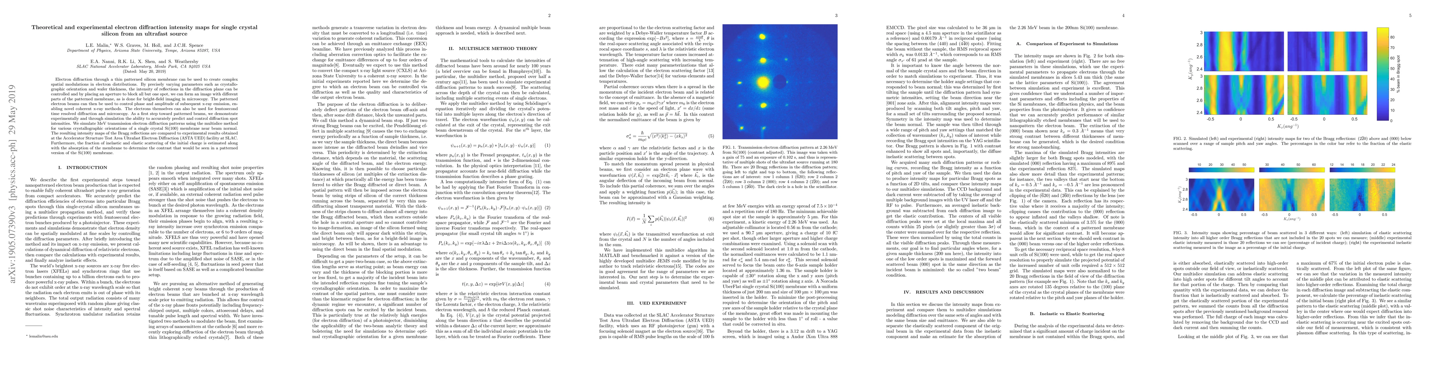
Electron diffraction through a thin patterned silicon membrane can be used to create complex spatial modulations in electron distributions by varying the intensity of different reflections using parameters such as crystallographic orientation and wafer thickness, then selecting specific spots in the diffraction plane using apertures. The patterned electron beams can be used to control phase and amplitude of subsequent x-ray emission, enabling novel coherent x-ray methods. The electrons themselves can also be used for femtosecond time resolved diffraction and microscopy. As a first step toward patterned beams, we demonstrate experimentally and through simulation the ability to accurately predict and control diffraction spot intensities. We simulate MeV transmission electron diffraction patterns using the multislice method for various crystallographic orientations of a single crystal Si(100) membrane near beam normal. The resulting intensity maps of the Bragg reflections are compared to experimental results obtained at the Accelerator Structure Test Area Ultrafast Electron Diffraction (ASTA UED) facility at SLAC. Furthermore, the fraction of inelastic and elastic scattering of the initial charge is estimated along with the absorption of the membrane to determine the contrast that would be seen in a patterned version of the Si(100) membrane.
AI Key Findings
Get AI-generated insights about this paper's methodology, results, significance, and more — seven facets brought into focus.
Impact
Paper Details
PDF Preview
Key Terms
Citation Network
Current paper (gray), citations (green), references (blue)
Display is limited for performance on very large graphs.
Discussion 0