This paper introduces a Thermally Adaptive Surface Microscopy capable of high-speed, 3D functional imaging of neuronal activity in living brains. The innovative design uses thermally tunable microlenses for optical sectioning and simultaneous recording of 3D fluorescence dynamics in zebrafish larval brains at a rate of 0.5 kHz.
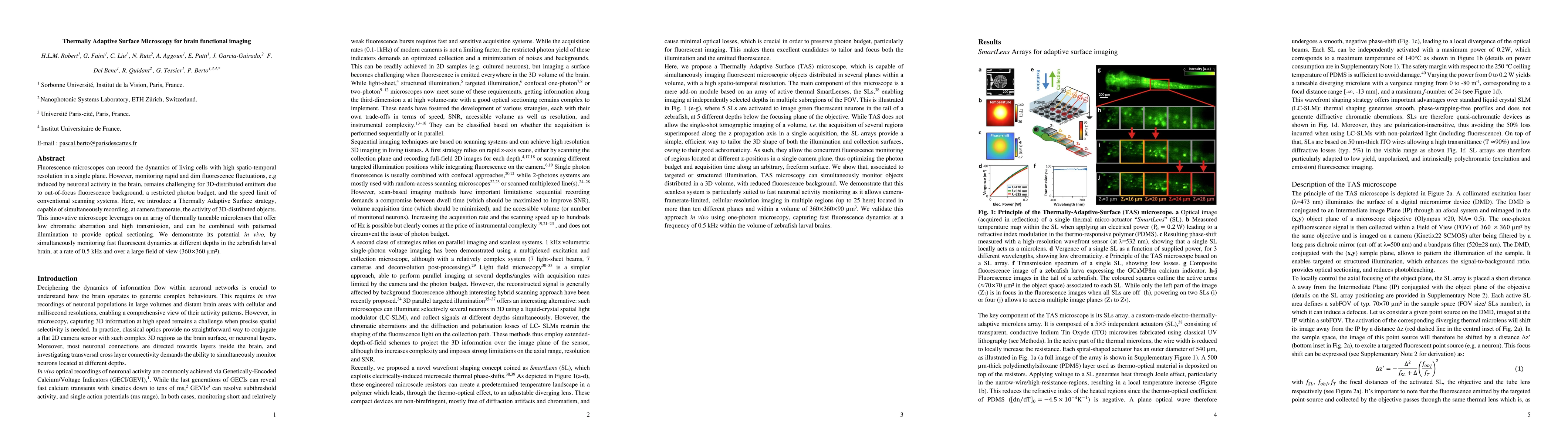
Fluorescence microscopes can record the dynamics of living cells with high spatio-temporal resolution in a single plane. However, monitoring rapid and dim fluorescence fluctuations, e.g induced by neuronal activity in the brain, remains challenging for 3D-distributed emitters due to out-of-focus fluorescence background, a restricted photon budget, and the speed limit of conventional scanning systems. Here, we introduce a Thermally Adaptive Surface strategy, capable of simultaneously recording, at camera framerate, the activity of 3D-distributed objects. This innovative microscope leverages on an array of thermally tuneable microlenses that offer low chromatic aberration and high transmission, and can be combined with patterned illumination to provide optical sectioning. We demonstrate its potential in vivo, by simultaneously monitoring fast fluorescent dynamics at different depths in the zebrafish larval brain, at a rate of 0.5 kHz and over a large field of view (360um x 360um).
Generated Sep 03, 2025
The research introduces a Thermally Adaptive Surface (TAS) microscopy strategy, utilizing an array of thermally tuneable microlenses for simultaneous 3D fluorescence imaging of distributed objects at camera framerate.
This research offers a novel, scanless method for parallel imaging of 3D fluorescent objects, particularly beneficial for studying complex cellular distributions in their natural, non-planar geometries, like in the brain.
The TAS microscope leverages thermo-optical effects for independent control of focus in multiple regions, providing a broadband, polarization-insensitive, refractive-based approach for shaping both illuminated and imaged surfaces.
Unlike existing scanless methods that primarily focus on optogenetics, this work presents a fluorescence imaging solution that requires wavefront modulation on both excitation and collection paths, offering a straightforward add-on to various microscopy techniques.
Current paper (gray), citations (green), references (blue)
Display is limited for performance on very large graphs.
Prevedel, Robert, Ferrer Ortas, Júlia, Kerr, Jason N D et al.
No citations found for this paper.
Comments (0)