This paper proposes a method to enhance 2D networks for 3D medical image segmentation by thickening 2D inputs with multiple slices, incorporating 3D contextual information. It uses early-stage multiplexing and slice-sensitive attention to mitigate information loss, achieving higher performance with lower latency for segmenting complex 3D structures like blood vessels in CT scans.
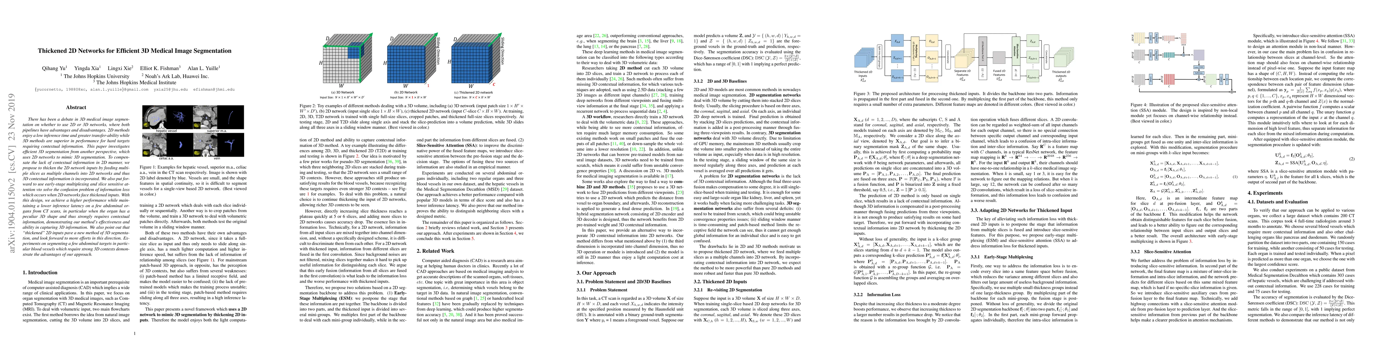
There has been a debate in 3D medical image segmentation on whether to use 2D or 3D networks, where both pipelines have advantages and disadvantages. 2D methods enjoy a low inference time and greater transfer-ability while 3D methods are superior in performance for hard targets requiring contextual information. This paper investigates efficient 3D segmentation from another perspective, which uses 2D networks to mimic 3D segmentation. To compensate the lack of contextual information in 2D manner, we propose to thicken the 2D network inputs by feeding multiple slices as multiple channels into 2D networks and thus 3D contextual information is incorporated. We also put forward to use early-stage multiplexing and slice sensitive attention to solve the confusion problem of information loss which occurs when 2D networks face thickened inputs. With this design, we achieve a higher performance while maintaining a lower inference latency on a few abdominal organs from CT scans, in particular when the organ has a peculiar 3D shape and thus strongly requires contextual information, demonstrating our method's effectiveness and ability in capturing 3D information. We also point out that "thickened" 2D inputs pave a new method of 3D segmentation, and look forward to more efforts in this direction. Experiments on segmenting a few abdominal targets in particular blood vessels which require strong 3D contexts demonstrate the advantages of our approach.
This graph shows how this paper is connected to other research. The current paper is shown in dark gray, papers that cite it are in green, and papers it references are in blue. You can:
• Hover over any node to see the paper title
• Click a node to view that paper's details
• Drag nodes to rearrange the graph
• Use the zoom controls (bottom right) to zoom in/out
Title | Authors | Year | Actions |
---|---|---|---|
Pixel level deep reinforcement learning for accurate and robust medical image segmentation
|
Yunxin Liu, Di Yuan, Zhenghua Xu, Yuefu Zhan, Hongwei Zhang, Jun Lu, Thomas Lukasiewicz
|
2025
|
View Paper |
Incorporating 3D Information in 2.5D Networks: Strategies for MR to CT Synthesis
|
Arthur Longuefosse, B. D. Senneville, G. Dournes, I. Benlala, P. Desbarats, F. Baldacci
|
2024
|
View Paper |
Assessing the Performance of U-Net in 3D Medical Image Segmentation
|
Khenaifer Fatma, Ilyes Benaissa, A. Zitouni, Baarir Zinne-eddine
|
2024
|
View Paper |
CSAM: A 2.5D Cross-Slice Attention Module for Anisotropic Volumetric Medical Image Segmentation
|
A. Hung, Haoxin Zheng, Kai Zhao, Xiaoxi Du, Kaifeng Pang, Qi Miao, Steven S. Raman, D. Terzopoulos, Kyunghyun Sung
|
2023
|
View Paper |
Evaluation of synthetically generated computed tomography for use in transcranial focused ultrasound procedures
|
Han Liu, Michelle K. Sigona, Thomas J. Manuel, L. Chen, Benoit M. Dawant, C. Caskey
|
2023
|
View Paper |
LSW‐Net: Lightweight Deep Neural Network Based on Small‐World properties for Spine MR Image Segmentation
|
Siyuan He, Qi Li, Xianda Li, Mengchao Zhang
|
2023
|
View Paper |
An efficient 3D reconstruction method based on WT-TV denoising for low-dose CT images
|
Yaoyao Liu, Chang Wang
|
2023
|
View Paper |
SDA-UNet: a hepatic vein segmentation network based on the spatial distribution and density awareness of blood vessels
|
Guoyu Tong, Huiyan Jiang, Yuan Yao
|
2023
|
View Paper |
Evaluation of Synthetically Generated CT for use in Transcranial Focused Ultrasound Procedures
|
Han Liu, Michelle K. Sigona, Thomas J. Manuel, L. Chen, B. Dawant, C. Caskey
|
2022
|
View Paper |
MRF-Net: A multi-branch residual fusion network for fast and accurate whole-brain MRI segmentation
|
Chong Wei, Yanwu Yang, Xutao Guo, Chenfei Ye, Haiyan Lv, Yang Xiang, Ting Ma
|
2022
|
View Paper |
FAST-AID Brain: Fast and Accurate Segmentation Tool using Artificial Intelligence Developed for Brain
|
Mostafa Mehdipour-Ghazi, Mads Nielsen
|
2022
|
View Paper |
Oral Cone Beam Computed Tomography Images Segmentation Based On Multi-view Fusion
|
Yunyao Jin, Qijun Zhao, Jun-ge Cheng
|
2022
|
View Paper |
Synthetic CT skull generation for transcranial MR imaging–guided focused ultrasound interventions with conditional adversarial networks
|
Han Liu, Michelle K. Sigona, Thomas J. Manuel, L. Chen, C. Caskey, B. Dawant
|
2022
|
View Paper |
A 2.5D semantic segmentation of the pancreas using attention guided dual context embedded U-Net
|
Jingyuan Li, Guanqun Liao, Wen-Kai Sun, Ji Sun, Tai Sheng, K. Zhu, K. M. Deneen, Yi Zhang
|
2022
|
View Paper |
CS-Net: Instance-aware cellular segmentation with hierarchical dimension-decomposed convolutions and slice-attentive learning
|
Jialin Peng, Zhengrong Luo
|
2021
|
View Paper |
Mixed 2D and 3D convolutional network with multi-scale context for lesion segmentation in breast DCE-MRI
|
Hongyu Wang, Jiaqi Cao, Jun Feng, Yilin Xie, Di Yang, Bao-ying Chen
|
2021
|
View Paper |
Recurrent Convolutional Neural Networks for 3D Mandible Segmentation in Computed Tomography
|
Bingjiang Qiu, Jiapan Guo, J. Kraeima, H. Glas, Weichuan Zhang, R. Borra, M. Witjes, P. V. van Ooijen
|
2021
|
View Paper |
GAU-Net: U-Net Based on Global Attention Mechanism for brain tumor segmentation
|
Xiuling Gan, Lidan Wang, Qi Chen, Yongjie Ge, Shukai Duan
|
2021
|
View Paper |
Automatic segmentation of ventricular volume by 3D ultrasonography in post haemorrhagic ventricular dilatation among preterm infants
|
L. C. Gontard, J. Pizarro, Borja Sanz-Peña, S. L. Lubián López, Isabel Benavente-Fernández
|
2021
|
View Paper |
Planar 3D Transfer Learning for End to End Unimodal MRI Unbalanced Data Segmentation
|
M. Kolarík, Radim Burget, C. Travieso-González, Jan Kocica
|
2020
|
View Paper |
Bridging 2D and 3D segmentation networks for computation-efficient volumetric medical image segmentation: An empirical study of 2.5D solutions
|
Yichi Zhang, Qingcheng Liao, Leiting Ding, Jicong Zhang
|
2020
|
View Paper |
Exploring Efficient Volumetric Medical Image Segmentation Using 2.5D Method: An Empirical Study
|
Yichi Zhang, Qingcheng Liao, Jicong Zhang
|
2020
|
View Paper |
Automatic Skull Stripping of Rat and Mouse Brain MRI Data Using U-Net
|
Li-Ming Hsu, Shuai Wang, Paridhi Ranadive, Woomi Ban, T. Chao, Shengdeng Song, Domenic H. Cerri, Lindsay R. Walton, Margaret A. Broadwater, Sung-Ho Lee, D. Shen, Y. Shih
|
2020
|
View Paper |
Conquering Data Variations in Resolution: A Slice-Aware Multi-Branch Decoder Network
|
Shuxin Wang, Shilei Cao, Zhizhong Chai, Dong Wei, Kai Ma, Liansheng Wang, Yefeng Zheng
|
2020
|
View Paper |
Deep learning to estimate the physical proportion of infected region of lung for COVID-19 pneumonia with CT image set
|
Wei Wu, Yu Shi, Xukun Li, Yukun Zhou, P. Du, Shuangzhi Lv, T. Liang, J. Sheng
|
2020
|
View Paper |
Distill DSM: Computationally efficient method for segmentation of medical imaging volumes
|
Harsh Maheshwari, Vidit Goel, Ramanathan Sethuraman, Debdoot Sheet
|
2021
|
View Paper |
TomoSAR 3D Reconstruction for Buildings Using Very Few Tracks of Observation: A Conditional Generative Adversarial Network Approach
|
Shihong Wang, Jiayi Guo, Yueting Zhang, Yuxin Hu, C. Ding, Yirong Wu
|
2021
|
View Paper |
Published: 2019-07-29
Published: 2022-02-08
Mykola Pechenizkiy, Shiwei Liu, Lu Yin, Qiao Xiao, Boqian Wu, Decebal Constantin Mocanu, Maurice Van Keulen, Elena Mocanu
Published: 2023-12-11
Comments (0)