Publication
Metrics
AI Quick Summary
This paper reports the first three-dimensional coherent X-ray diffraction imaging (cryo-CDI) of a whole, frozen-hydrated cell, revealing its surface and internal morphology at a resolution of ~75-100 nm. The study forecasts improved spatial resolutions in the future as coherent X-ray flux and detector technology advance.
Paper Preview
Abstract
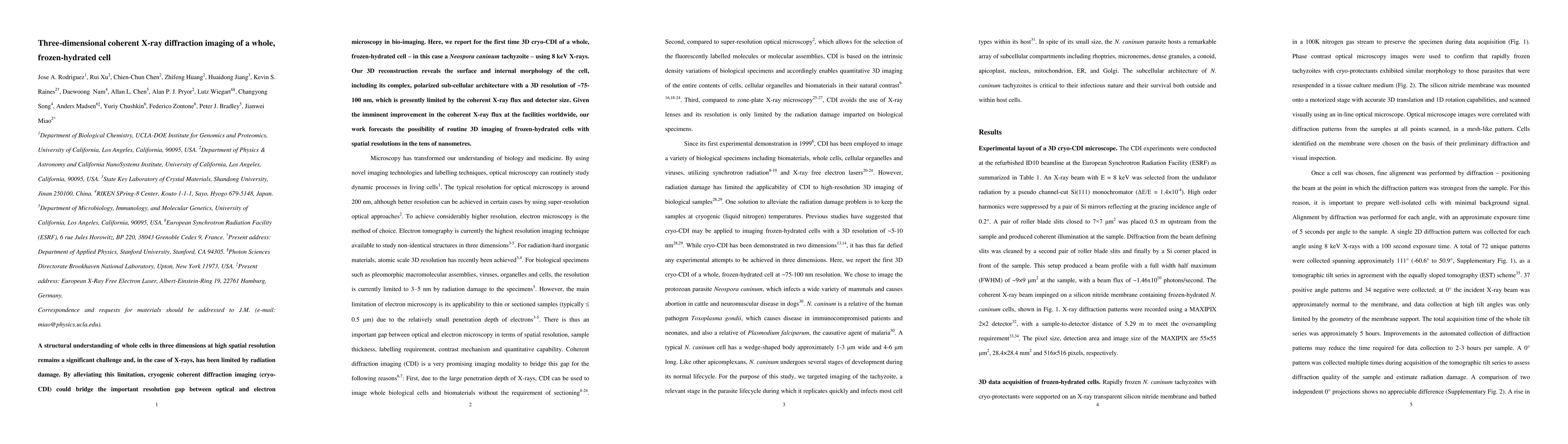
A structural understanding of whole cells in three dimensions at high spatial resolution remains a significant challenge and, in the case of X-rays, has been limited by radiation damage. By alleviating this limitation, cryogenic coherent diffraction imaging (cryo-CDI) could bridge the important resolution gap between optical and electron microscopy in bio-imaging. Here, we report for the first time 3D cryo-CDI of a whole, frozen-hydrated cell - in this case a Neospora caninum tachyzoite - using 8 keV X-rays. Our 3D reconstruction reveals the surface and internal morphology of the cell, including its complex, polarized sub-cellular architecture with a 3D resolution of ~75-100 nm, which is presently limited by the coherent X-ray flux and detector size. Given the imminent improvement in the coherent X-ray flux at the facilities worldwide, our work forecasts the possibility of routine 3D imaging of frozen-hydrated cells with spatial resolutions in the tens of nanometres.
AI Key Findings
Get AI-generated insights about this paper's methodology, results, significance, and more — seven facets brought into focus.
Impact
Paper Details
PDF Preview
Key Terms
Citation Network
Current paper (gray), citations (green), references (blue)
Display is limited for performance on very large graphs.
Discussion 0