Three-dimensional Generative Adversarial Nets for Unsupervised Metal Artifact Reduction
Publication
Metrics
AI Quick Summary
This paper proposes an unsupervised three-dimensional generative adversarial network (GAN) for reducing metal artifacts in CT images, particularly from dental fillings. The method effectively reduces artifacts and recovers missing voxels while preserving anatomical features, as demonstrated by experiments on 915 clinical CT volumes.
Paper Preview
Abstract
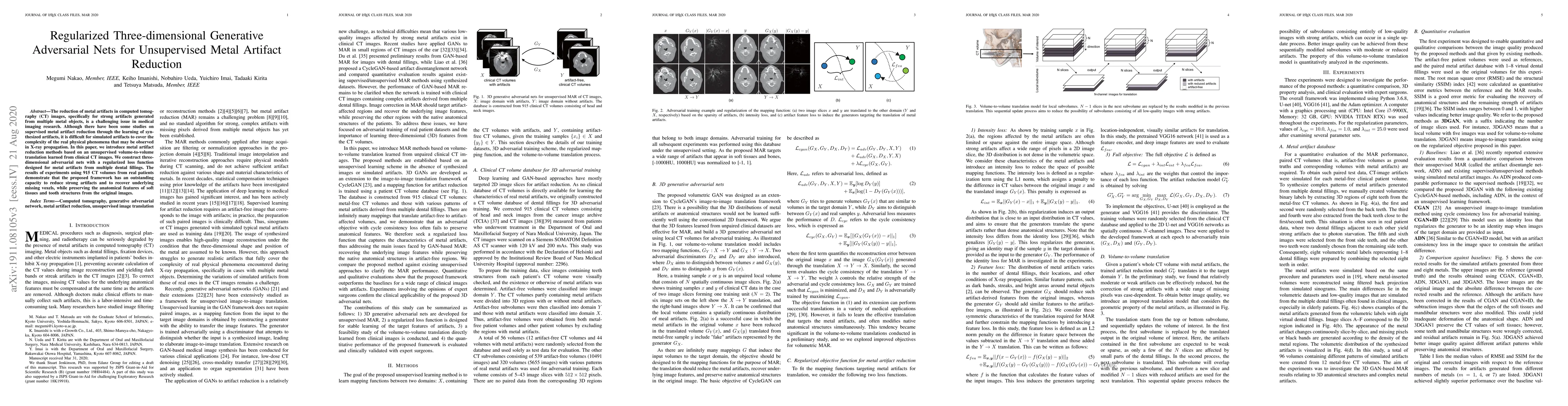
The reduction of metal artifacts in computed tomography (CT) images, specifically for strong artifacts generated from multiple metal objects, is a challenging issue in medical imaging research. Although there have been some studies on supervised metal artifact reduction through the learning of synthesized artifacts, it is difficult for simulated artifacts to cover the complexity of the real physical phenomena that may be observed in X-ray propagation. In this paper, we introduce metal artifact reduction methods based on an unsupervised volume-to-volume translation learned from clinical CT images. We construct three-dimensional adversarial nets with a regularized loss function designed for metal artifacts from multiple dental fillings. The results of experiments using 915 CT volumes from real patients demonstrate that the proposed framework has an outstanding capacity to reduce strong artifacts and to recover underlying missing voxels, while preserving the anatomical features of soft tissues and tooth structures from the original images.
AI Key Findings
Get AI-generated insights about this paper's methodology, results, significance, and more — seven facets brought into focus.
Impact
Paper Details
PDF Preview
Key Terms
Citation Network
Current paper (gray), citations (green), references (blue)
Display is limited for performance on very large graphs.
Discussion 0