This paper presents an x-ray microtomographic technique for imaging the 3D structure of the human cerebral cortex, enhanced by staining neural cells with metal compounds to improve contrast and reveal the microarchitecture of gray and white matter regions in the frontal cortex.
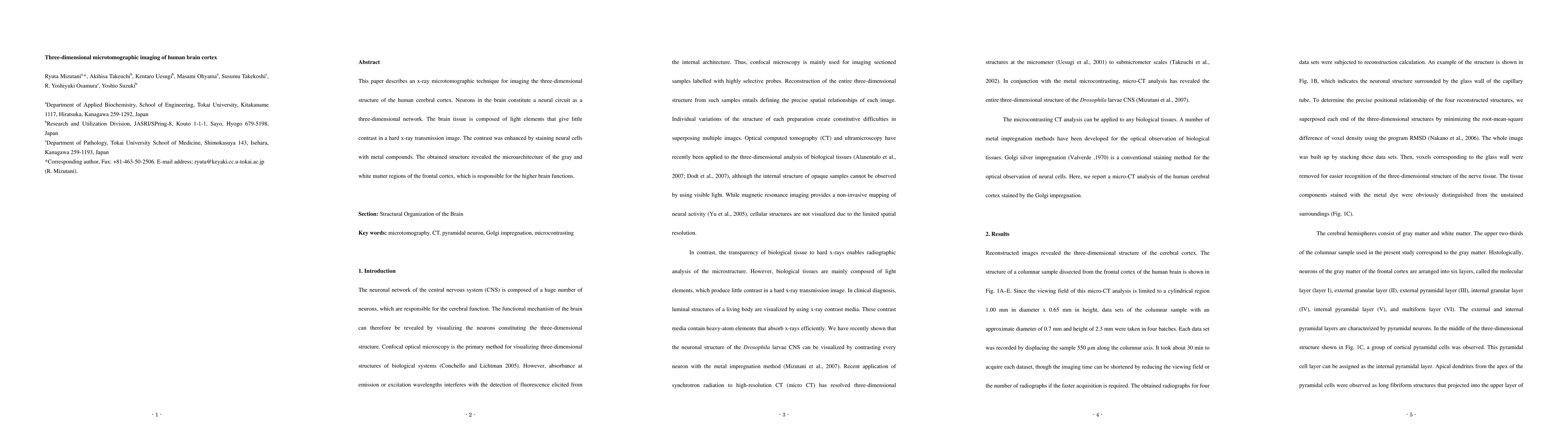
This paper describes an x-ray microtomographic technique for imaging the three-dimensional structure of the human cerebral cortex. Neurons in the brain constitute a neural circuit as a three-dimensional network. The brain tissue is composed of light elements that give little contrast in a hard x-ray transmission image. The contrast was enhanced by staining neural cells with metal compounds. The obtained structure revealed the microarchitecture of the gray and white matter regions of the frontal cortex, which is responsible for the higher brain functions.
Get AI-generated insights about this paper's methodology, results, and significance.
Current paper (gray), citations (green), references (blue)
Michael D. Brown, Christos Strydis, Bastian S. Generowicz et al.
Title | Authors | Year | Actions |
---|---|---|---|
Three-Dimensional Microscopic Characteristics of the Human Uterine Cervix Evaluated by Microtomography
|
A. P. Matos, O. L. Aranda, Edson Marchiori, Alessandra Silveira Machado, Adriana José Da Penha Moreira, H. Werner, E. Araujo Júnior, R. Granese, G. Calagna, P. T. Castro
|
2025
|
View Paper |
Three-Dimensional Cell Culture Micro-CT Visualization within Collagen Scaffolds in an Aqueous Environment
|
S. Tkachev, N. Chepelova, G. Galechyan, Boris Ershov, Danila Golub, E. Popova, A. Antoshin, Aliia Giliazova, Sergei Voloshin, Yu. I. Efremov, Elena Istranova, Peter Timashev
|
2024
|
View Paper |
Virtual histology of an entire mouse brain from formalin fixation to paraffin embedding. Part 1: Data acquisition, anatomical feature segmentation, tracking global volume and density changes
|
Griffin Rodgers, W. Kuo, G. Schulz, M. Scheel, Alexandra Migga, C. Bikis, C. Tanner, V. Kurtcuoglu, T. Weitkamp, B. Müller
|
2021
|
View Paper |
Brain capillary structures of schizophrenia cases and controls show a correlation with their neuron structures
|
R. Saiga, M. Uesugi, A. Takeuchi, K. Uesugi, Yoshio Suzuki, S. Takekoshi, C. Inomoto, Naoya Nakamura, Youta Torii, I. Kushima, S. Iritani, N. Ozaki, Kenichi Oshima, M. Itokawa, Makoto Arai, R. Mizutani
|
2021
|
View Paper |
A combinatorial method to visualize the neuronal network in the mouse spinal cord: combination of a modified Golgi-Cox method and synchrotron radiation micro-computed tomography
|
Liyuan Jiang, Yong Cao, Xianzhen Yin, Shuangfei Ni, Miao Li, Chengjun Li, Zixiang Luo, Hongbin Lu, Jianzhong Hu
|
2021
|
View Paper |
Visualization of different anatomical parts of the enucleated human eye using X-ray micro-CT imaging.
|
S. Tkachev, B. Mitrin, N. Karnaukhov, E. Sadyrin, M. Voloshin, A. Maksimov, A. Goncharova, E. Lukbanova, E. Zaikina, A. V. Volkova, D. V. Khodakova, M. V. Mindar, M. Yengibarian, T. Protasova, S. O. Kit, A. Ermakov, S. Chapek, M. Tkacheva
|
2020
|
View Paper |
Structural diverseness of neurons between brain areas and between cases
|
R. Mizutani, R. Saiga, Yoshiro Yamamoto, M. Uesugi, A. Takeuchi, K. Uesugi, Y. Terada, Yoshio Suzuki, V. De Andrade, F. De Carlo, S. Takekoshi, C. Inomoto, Naoya Nakamura, Youta Torii, I. Kushima, S. Iritani, N. Ozaki, Kenichi Oshima, M. Itokawa, Makoto Arai
|
2020
|
View Paper |
A synchrotron X-ray imaging strategy to map large animal brains
|
A. Chin, S. Yang, Hsiang-Hsin Chen, Min-Tsang Li, T. Lee, Ying-Jie Chen, Ting-Kuo Lee, C. Petibois, Xiaoqing Cai, C. Low, F. Tan, Alvin Teo, E. Tok, Edwin B. L. Ong, Yen-Yin Lin, I‐Jin Lin, Yi-Chi Tseng, N. Chen, C. Shih, Jae-Hong Lim, Jun Lim, J. Je, Y. Kohmura, T. Ishikawa, G. Margaritondo, A. Chiang, Y. Hwu
|
2020
|
View Paper |
Cutting‐edge morphological studies of post‐mortem brains of patients with schizophrenia and potential applications of X‐ray nanotomography (nano‐CT)
|
M. Itokawa, Kenichi Oshima, Makoto Arai, Yota Torii, I. Kushima, S. Iritani, N. Ozaki, R. Saiga, R. Mizutani
|
2019
|
View Paper |
Comparative analysis of sample preparation protocols of soft biological tissues for morphometric studies using synchrotron-based X-ray microtomography.
|
C. S. Dias, Dionísio Pedro Amorim Neto, G. Baraldi, M. Fonseca
|
2019
|
View Paper |
Nanometer-Scale Structures of Neurons Differ Between Individuals and Those Differences Become Extraordinary in Schizophrenia
|
R. Saiga, V. Andrade, F. Carlo, A. Takeuchi, K. Uesugi, Y. Terada, Yoshio Suzuki, S. Takekoshi, C. Inomoto, Naoya Nakamura, Youta Torii, I. Kushima, S. Iritani, N. Ozaki, Kenichi Oshima, M. Itokawa, Makoto Arai, R. Mizutani
|
2019
|
View Paper |
High-resolution synchrotron-based X-ray microtomography as a tool to unveil the three-dimensional neuronal architecture of the brain
|
M. Fonseca, B. Araujo, C. S. Dias, N. Archilha, Dionísio Pedro Amorim Neto, E. Cavalheiro, H. Westfahl, Antônio José Roque da Silva, K. Franchini
|
2018
|
View Paper |
Preparation of soft tissue samples for synchrotron radiation microtomography and nanotomography
|
R. Mizutani, R. Saiga
|
2018
|
View Paper |
Three-dimensional alteration of neurites in schizophrenia
|
R. Mizutani, R. Saiga, A. Takeuchi, K. Uesugi, Y. Terada, Yoshio Suzuki, V. De Andrade, F. De Carlo, S. Takekoshi, C. Inomoto, Naoya Nakamura, I. Kushima, S. Iritani, N. Ozaki, S. Ide, K. Ikeda, Kenichi Oshima, M. Itokawa, Makoto Arai
|
2018
|
View Paper |
Brain tissue fingerprints of schizophrenia and control individuals
|
R. Mizutani, R. Saiga, A. Takeuchi, K. Uesugi, Y. Terada, Yoshio Suzuki, V. Andrade, F. Carlo, S. Takekoshi, C. Inomoto, Naoya Nakamura, I. Kushima, S. Iritani, N. Ozaki, S. Ide, K. Ikeda, Kenichi Oshima, M. Itokawa, Makoto Arai
|
2018
|
View Paper |
Submicrometer tomographic resolution examined using a micro-fabricated test object.
|
R. Mizutani, A. Takeuchi, R. Osamura, S. Takekoshi, K. Uesugi, Yoshio Suzuki
|
2016
|
View Paper |
A method for estimating spatial resolution of real image in the Fourier domain
|
R. Mizutani, R. Saiga, S. Takekoshi, C. Inomoto, Naoya Nakamura, M. Itokawa, Makoto Arai, Kenichi Oshima, A. Takeuchi, K. Uesugi, Y. Terada, Yoshio Suzuki
|
2016
|
View Paper |
Technique: Imaging Earliest Tooth Development in 3D Using a Silver‐Based Tissue Contrast Agent
|
Muhammad Raj, Martin A. Prusinkiewicz, D. Cooper, B. George, M. A. Webb, J. Boughner
|
2014
|
View Paper |
Upgraded X-ray topography and microtomography beamline at the Kurchatov synchrotron radiation source
|
R. Senin, A. S. Khlebnikov, A. E. Vyazovetskova, I. Blinov, A. O. Golubitskii, I. Kazakov, A. A. Vorob’ev, A. Buzmakov, V. Asadchikov, V. Shishkov, É. Mukhamedzhanov, M. Kovalchuk
|
2013
|
View Paper |
X-ray microtomography in biology.
|
R. Mizutani, Yoshio Suzuki
|
2012
|
View Paper |
Microtomographic analysis of neuronal circuits of human brain.
|
R. Mizutani, A. Takeuchi, K. Uesugi, S. Takekoshi, R. Osamura, Yoshio Suzuki
|
2010
|
View Paper |
[Current concepts for experimental micro-CT in small animals].
|
S. Bag, S. J. Schambach, H. Boll, L. Schilling, C. Groden, M. Brockmann
|
2010
|
View Paper |
Three-dimensional cellular and subcellular structures of human brain tissue determined by microtomography
|
R. Mizutani, A. Takeuchi, S. Takekoshi, R. Osamura, K. Uesugi, Yoshio Suzuki
|
2009
|
View Paper |
X-ray microtomographic imaging of three-dimensional structure of soft tissues.
|
R. Mizutani, A. Takeuchi, K. Uesugi, S. Takekoshi, R. Osamura, Yoshio Suzuki
|
2008
|
View Paper |
Evaluation of the improved three-dimensional resolution of a synchrotron radiation computed tomograph using a micro-fabricated test pattern.
|
R. Mizutani, A. Takeuchi, K. Uesugi, Yoshio Suzuki
|
2008
|
View Paper |
Hard X-ray microtomography for virtual histology of the brain with cellular resolution
|
2021
|
View Paper | |
Comparação de parâmetros e protocolos de aquisição e reconstrução de imagens de microtomografia para o diagnóstico de lesões de cárie incipientes : Comparison of acquisition and reconstruction parameters and protocols of microtomography images for the diagnosis of incipient caries lesions
|
Karla Rovaris da Silva
|
2017
|
View Paper |
Unveiling 3 D Biological Structures by X-ray Microtomography
|
R. Mizutani, A. Takeuchi, K. Uesugi, S. Takekoshi, R. Osamura, Y. Suzuki
|
2011
|
View Paper |
eview-ray microtomography in biology
|
Y. Mizutania, Yoshio Suzukib
|
2011
|
View Paper |
Application of micro-CT in small animal imaging.
|
S. J. Schambach, S. Bag, L. Schilling, C. Groden, M. Brockmann
|
2010
|
View Paper |
Unveiling 3D Biological Structures by X-ray Microtomography
|
R. Mizutani, A. Takeuchi, K. Uesugi, S. Takekoshi, Y. Suzuki
|
2010
|
View Paper |
Comments (0)