Three-dimensional Segmentation of the Scoliotic Spine from MRI using Unsupervised Volume-based MR-CT Synthesis
Publication
Metrics
AI Quick Summary
This paper presents an unsupervised method for segmenting scoliotic spines from MRI using unpaired volume-to-volume MR-CT synthesis via a 3D CycleGAN model. The synthesized CT volumes are then thresholded for vertebral bone segmentation, achieving a mean error of 3.41 ± 1.06 mm.
Paper Preview
Abstract
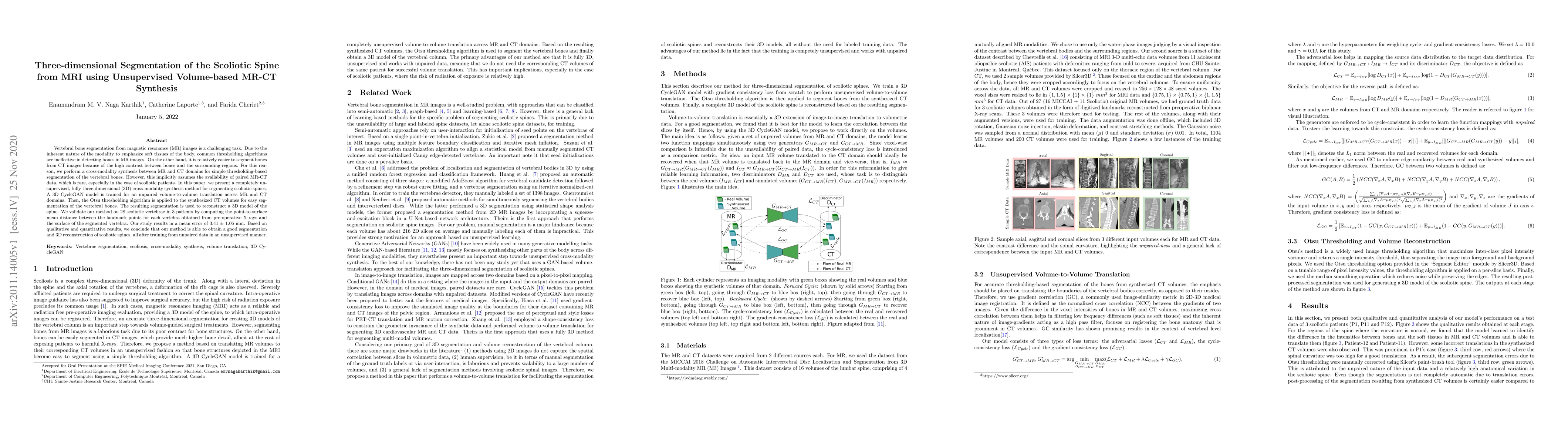
Vertebral bone segmentation from magnetic resonance (MR) images is a challenging task. Due to the inherent nature of the modality to emphasize soft tissues of the body, common thresholding algorithms are ineffective in detecting bones in MR images. On the other hand, it is relatively easier to segment bones from CT images because of the high contrast between bones and the surrounding regions. For this reason, we perform a cross-modality synthesis between MR and CT domains for simple thresholding-based segmentation of the vertebral bones. However, this implicitly assumes the availability of paired MR-CT data, which is rare, especially in the case of scoliotic patients. In this paper, we present a completely unsupervised, fully three-dimensional (3D) cross-modality synthesis method for segmenting scoliotic spines. A 3D CycleGAN model is trained for an unpaired volume-to-volume translation across MR and CT domains. Then, the Otsu thresholding algorithm is applied to the synthesized CT volumes for easy segmentation of the vertebral bones. The resulting segmentation is used to reconstruct a 3D model of the spine. We validate our method on 28 scoliotic vertebrae in 3 patients by computing the point-to-surface mean distance between the landmark points for each vertebra obtained from pre-operative X-rays and the surface of the segmented vertebra. Our study results in a mean error of 3.41 $\pm$ 1.06 mm. Based on qualitative and quantitative results, we conclude that our method is able to obtain a good segmentation and 3D reconstruction of scoliotic spines, all after training from unpaired data in an unsupervised manner.
AI Key Findings
Get AI-generated insights about this paper's methodology, results, significance, and more — seven facets brought into focus.
Impact
Paper Details
Authors
PDF Preview
Key Terms
Citation Network
Current paper (gray), citations (green), references (blue)
Display is limited for performance on very large graphs.
Discussion 0