Three-dimensional structural and compositional inhomogeneity in zeolites unraveled by low-dose electron ptychography
Publication
Metrics
AI Quick Summary
This study utilizes low-dose electron ptychography based on 4D-STEM to achieve ultrahigh lateral and depth resolution in zeolites, revealing three-dimensional structural and compositional inhomogeneity, including individual oxygen atoms and adsorbed molecules, and O vacancies. This method provides a comprehensive understanding of zeolite intergrowths.
Paper Preview
Abstract
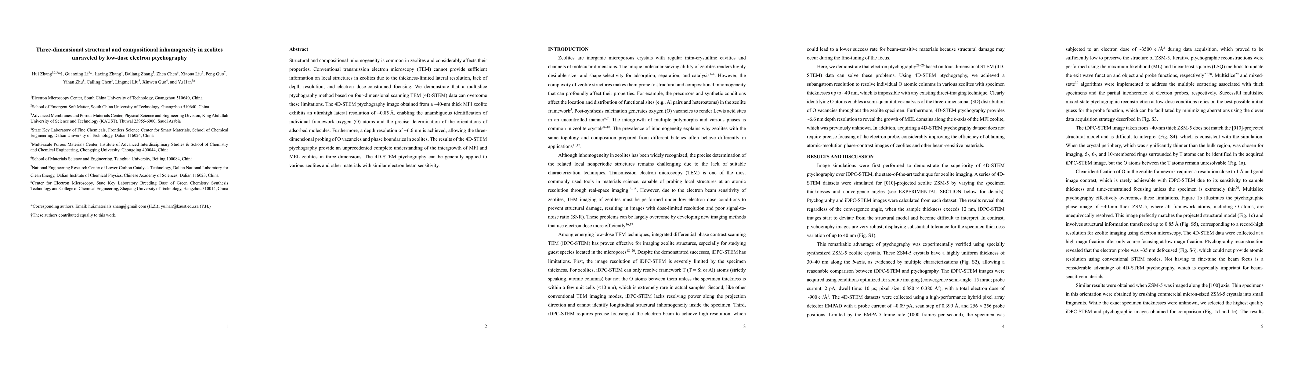
Structural and compositional inhomogeneity is common in zeolites and considerably affects their properties. Conventional transmission electron microscopy (TEM) cannot provide sufficient information on local structures in zeolites due to the thickness-limited lateral resolution, lack of depth resolution, and electron dose-constrained focusing. We demonstrate that a multislice ptychography method based on four-dimensional scanning TEM (4D-STEM) data can overcome these limitations. The 4D-STEM ptychography image obtained from a ~40-nm thick MFI zeolite exhibits an ultrahigh lateral resolution of ~0.85 {\AA}, enabling the unambiguous identification of individual framework oxygen (O) atoms and the precise determination of the orientations of adsorbed molecules. Furthermore, a depth resolution of ~6.6 nm is achieved, allowing the three-dimensional probing of O vacancies and phase boundaries in zeolites. The results of the 4D-STEM ptychography provide an unprecedented complete understanding of the intergrowth of MFI and MEL zeolites in three dimensions. The 4D-STEM ptychography can be generally applied to various zeolites and other materials with similar electron beam sensitivity.
AI Key Findings
Get AI-generated insights about this paper's methodology, results, significance, and more — seven facets brought into focus.
Impact
Paper Details
Authors
PDF Preview
Key Terms
Citation Network
Current paper (gray), citations (green), references (blue)
Display is limited for performance on very large graphs.
Discussion 0